Review
. 2019 Mar 26;12(1):141.
doi: 10.1186/s13071-019-3390-2.

Carrion's disease: more than a neglected disease

Affiliations
Review

Carrion's disease: more than a neglected disease

Meritxell Garcia-Quintanilla et al. Parasit Vectors. .

Abstract

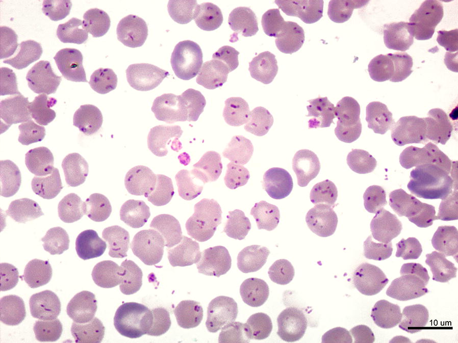
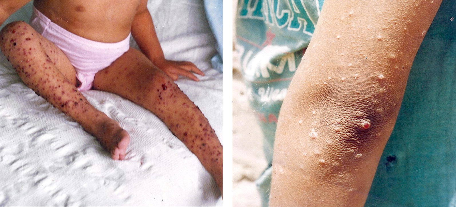
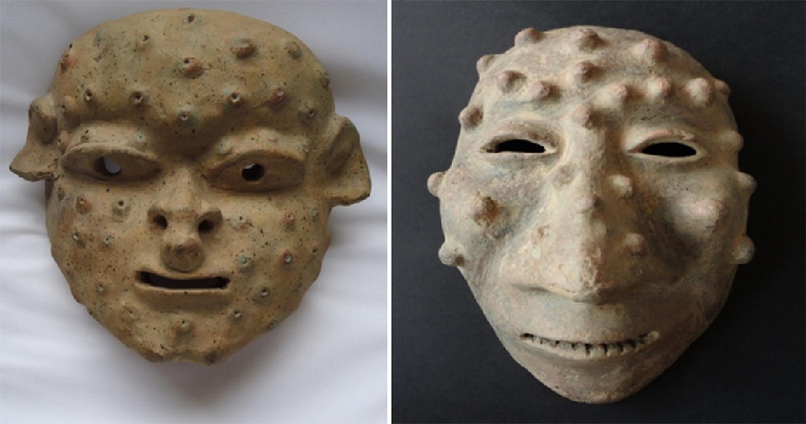
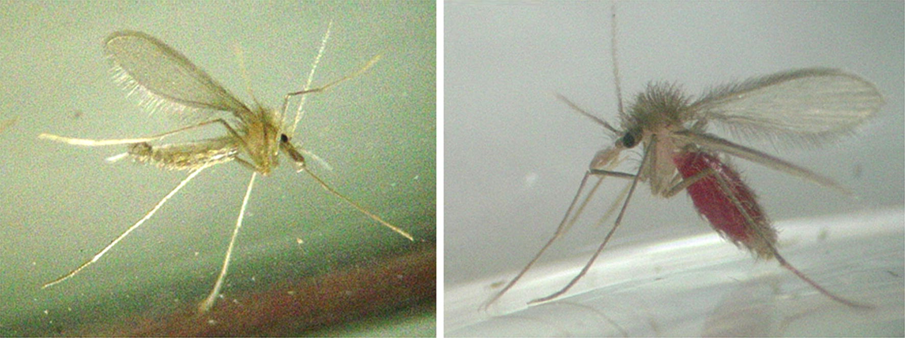
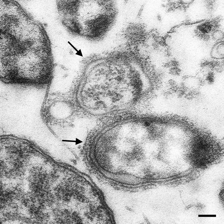
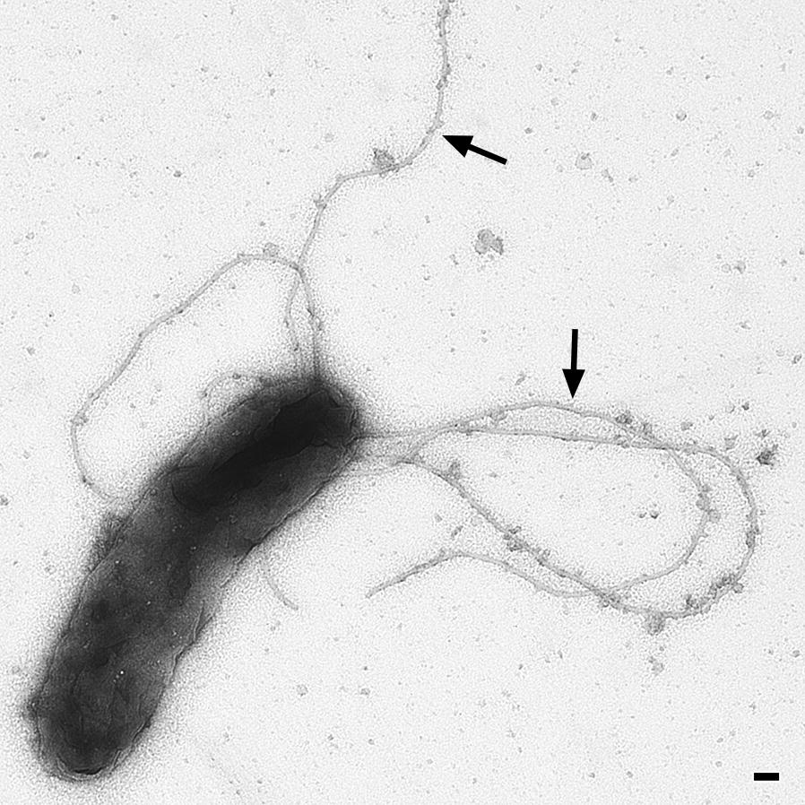
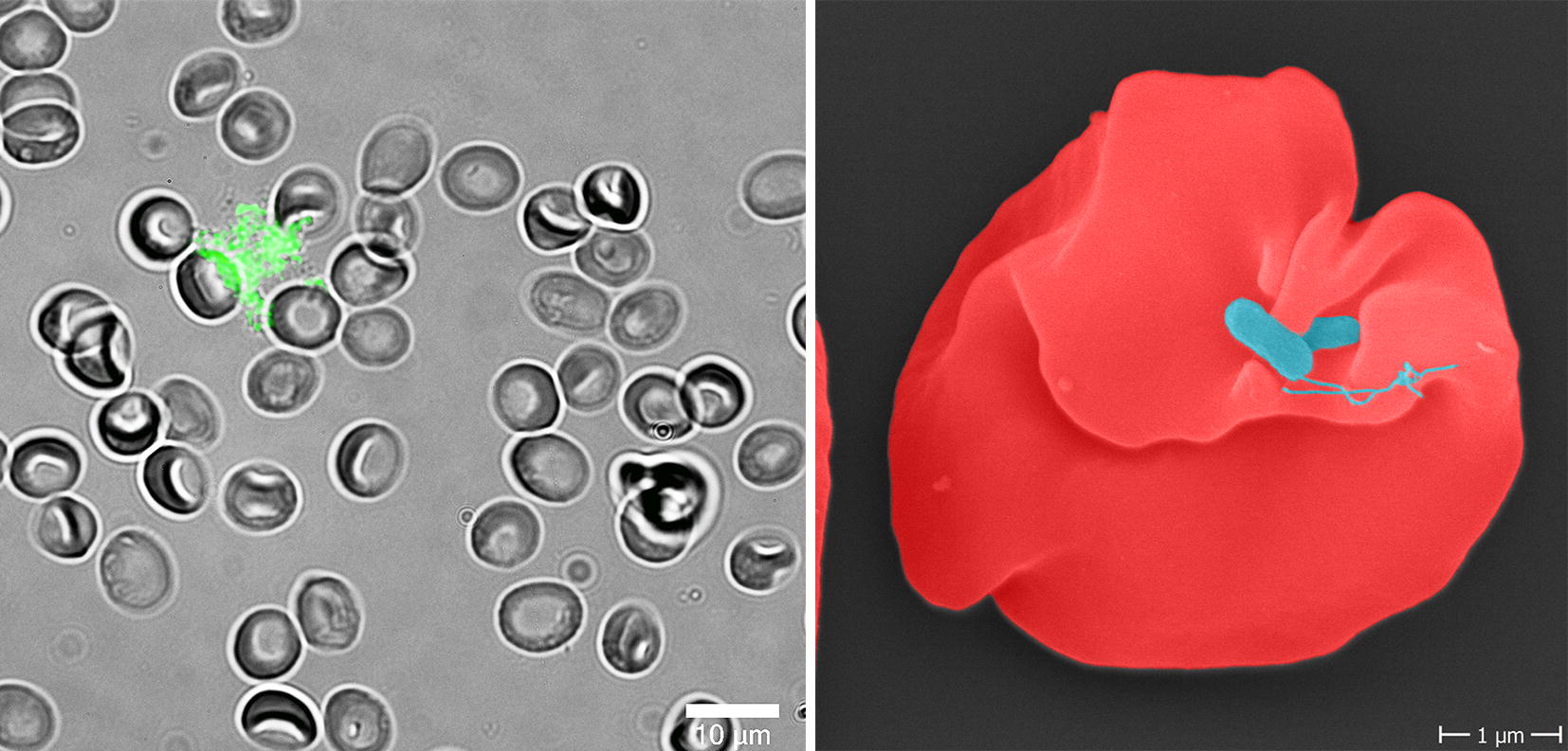
Infections with Bartonella bacilliformis result in Carrion's disease in humans. In the first phase of infection, the pathogen causes a hemolytic fever ("Oroya fever") with case-fatality rates as high as ~90% in untreated patients, followed by a chronical phase resulting in angiogenic skin lesions ("verruga peruana"). Bartonella bacilliformis is endemic to South American Andean valleys and is transmitted via sand flies (Lutzomyia spp.). Humans are the only known reservoir for this old disease and therefore no animal infection model is available. In the present review, we provide the current knowledge on B. bacilliformis and its pathogenicity factors, vectors, possible unknown reservoirs, established and potential infection models and immunological aspects of the disease.

Keywords: Bartonella bacilliformis; Carrion’s disease; Lutzomyia; Neglected tropical disease; South America; Vector-borne disease.


Conflict of interest statement

The authors declare that they have no competing interests.

Figures

Fig. 1
Fig. 1
Overwhelming parasitism of erythrocytes by B. bacilliformis. Giemsa-stained blood smear from a patient with Oroya fever, showing parasitism of all erythrocytes, with bacillary and coccoid forms of B. bacilliformis. Scale-bar: 10 µm (courtesy of P. Ventosilla and M. Montes, Universidad Peruana Cayetano Heredia, Lima, Peru)
Fig. 2
Fig. 2
Patients with Verruga peruana caused by B. bacilliformis. Left: 9-year-old girl with numerous bleeding verrugas on her legs; Huaraz, Ancash, 1993. Right: 17-year-old girl (facing left) showing multiple verrugas close to her left elbow; a single verruga has broken the overlying epidermis, and may later bleed; Huari, Ancash, 2002 (courtesy of C. Maguiña, Universidad Peruana Cayetano Heredia, Lima, Peru)
Fig. 3
Fig. 3
Ceramic masks (400 B.C.–400 A.D.). Two masks discovered in Ecuador displaying the facial symptoms of verruga peruana. Citation: Sotomayor-Tribín HA. Pensamiento analógico mítico en la interpretación del arte prehispánico de interés para la arqueomedicina y la paleopatología. Repert Med Cir. 2016;25:50–71 [94]. With permission of Elsevier
Fig. 4
Fig. 4
Adult Lutzomyia verrucarum sand flies. Left: male. Right: blood-fed female. Colony-bred adults. Length of each between 2 and 3 mm (courtesy of E. Pérez, Universidad Peruana Cayetano Heredia, Lima, Peru)
Fig. 5
Fig. 5
Bartonella bacilliformis adhesin A (BbadA) expressed on the surface of B. bacilliformis. Electron microscopy of B. bacilliformis ATCC 35686 (grown for four days at 28 °C in Bartonella liquid medium [95]). Arrows indicate the presumptive BbadA expression on the bacterial surface. Scale-bar: 100 nm (courtesy of M. Schaller and B. Fehrenbacher, Eberhard Karls-University, Tuebingen, Germany)
Fig. 6
Fig. 6
Flagella of B. bacilliformis. Electron microscopy of B. bacilliformis ATCC 35686 (grown for four days at 28 °C in Bartonella liquid medium [95]). Arrows indicate the presumptive BbadA expression on the bacterial surface. Scale-bar: 100 nm (courtesy of M. Schaller and B. Fehrenbacher, Eberhard Karls-University, Tuebingen, Germany)
Fig. 7
Fig. 7
Human erythrocytes infected with B. bacilliformis. Left: Fluorescence microscopy of human erythrocytes infected with GFP-expressing B. bacilliformis ATCC 35686 (6 h). Note the deformation of the erythrocyte cell surface (Aepfelbacher and Kempf, 2018). Scale-bar: 10 µm. Right: Scanning electron microscopy of infected human erythrocytes (24 h). Note the deformation of the erythrocyte. Scale-bar: 1 µm (courtesy of C. Sittmann, Goethe University, Frankfurt am Main, Germany and K. Hipp, Max Planck-Institute for developmental Biology, Tuebingen, Germany)

Similar articles

Cited by

References

    1. Amano Y, Rumbea J, Knobloch J, Olson J, Kron M. Bartonellosis in Ecuador: serosurvey and current status of cutaneous verrucous disease. Am J Trop Med Hyg. 1997;57:174–179. - PubMed
    1. Lydy SL, Lascano MS, Garcia-Perez JE, Williams-Newkirk AJ, Grijalva MJ. Seroprevalence and risk factors for infection with Bartonella bacilliformis in Loja Province, Ecuador. Emerg Microbes Infect. 2018;7:115. - PMC - PubMed
    1. Gomes C, Ruiz J. Carrion’s disease: the sound of silence. Clin Microbiol Rev. 2018;31:e00056. - PMC - PubMed
    1. Maguina C, Garcia PJ, Gotuzzo E, Cordero L, Spach DH. Bartonellosis (Carrionʼs disease) in the modern era. Clinical Infect Dis. 2001;33:772–779. - PubMed
    1. Minnick MF, Anderson BE, Lima A, Battisti JM, Lawyer PG, Birtles RJ. Oroya fever and verruga peruana: bartonelloses unique to South America. PLoS Negl Trop Dis. 2014;8:e2919. - PMC - PubMed

MeSH terms

LinkOut - more resources