. 2019 Mar 25;2(2):e201800218.
doi: 10.26508/lsa.201800218. Print 2019 Apr.

Location-dependent maintenance of intrinsic susceptibility to mTORC1-driven tumorigenesis

Affiliations

Location-dependent maintenance of intrinsic susceptibility to mTORC1-driven tumorigenesis

Gabrielle V Rushing et al. Life Sci Alliance. .

Abstract

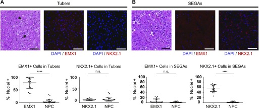
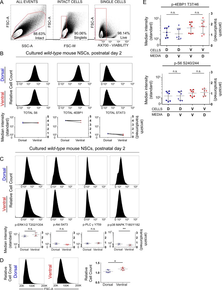
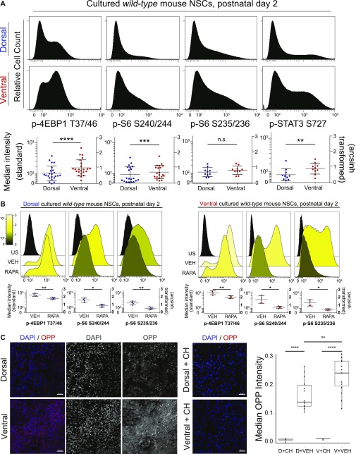
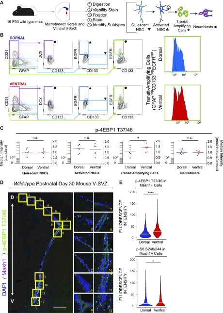
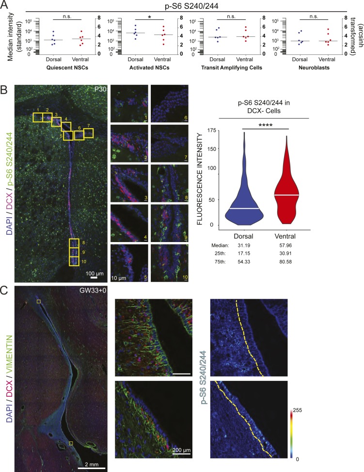
Neural stem/progenitor cells (NSPCs) of the ventricular-subventricular zone (V-SVZ) are candidate cells of origin for many brain tumors. However, whether NSPCs in different locations within the V-SVZ differ in susceptibility to tumorigenic mutations is unknown. Here, single-cell measurements of signal transduction intermediates in the mechanistic target of rapamycin complex 1 (mTORC1) pathway reveal that ventral NSPCs have higher levels of signaling than dorsal NSPCs. These features are linked with differences in mTORC1-driven disease severity: introduction of a pathognomonic Tsc2 mutation only results in formation of tumor-like masses from the ventral V-SVZ. We propose a direct link between location-dependent intrinsic growth properties imbued by mTORC1 and predisposition to tumor development.


Conflict of interest statement

JM Irish is a co-founder and board member at Cytobank Inc. and received research support from Incyte Corp., Janssen, and Pharmacyclics. The other authors declare no conflict of interest.

Figures

Figure 1.
Figure 1.. Transcription factors defining dorsal or ventral identity are differentially expressed in dorsal and ventral hamartomas in TSC.
(A) Representative fields from tilescans of human TSC cortical tubers stained with hematoxylin and eosin (left) or for DAPI (blue) and EMX1 (center, red) or NKX2.1 (right, red). Note the enlarged balloon cells (arrows) typical of tubers. Quantification of % positive nuclei for each factor is shown below. EMX1 is abundantly expressed (P < 0.0001 versus NPC), whereas NKX2.1 is not (P = 0.5809). N = 4 tubers, each dot = 1 region of interest (ROI), 4–5 ROIs/tuber. (B) Representative fields from tilescans of human SEGA tumors stained with hematoxylin and eosin (left) or for DAPI (blue) and EMX1 (center, red) or NKX2.1 (right, red). Quantification of positive nuclei is shown below, as in (A). In these ventral tumors, EMX1 is not widely expressed (P = 0.3373), but NKX2.1 is abundant (P < 0.0001). N = 4 SEGAs, each dot = 1 ROI, 5 ROIs/SEGA. Mann–Whitney tests were used. All scale bars = 100 μm.
Figure S1.
Figure S1.. V-SVZ cultures express transcription factors expected for region of origin.
(A) Cartoon schematic of culture preparation. (B) Representative confocal images of P2 V-SVZ cultures stained for DAPI (blue), NKX2.1 (red), and PAX6 (green) protein. PAX6 and NKX2.1 staining are shown in grayscale to the right. Quantification of positive nuclei in dorsal and ventral cultures is displayed as box and whisker plots to the right; bars, median + 25th and 75th percentile ± SD. Scale bars, 50 μm. n = 6 mice, 3 ROIs per mouse. Paired t tests, P < 0.001 (Pax6), P = 0.007 (Nkx2.1). (C) Graph showing transcript abundance for the transcription factors NKX2.1, NKX6.2, and PAX6 from P2 V-SVZ cultures. Ventral ΔCT is subtracted from Dorsal ΔCT; therefore, transcripts higher in dorsal samples are above 0 and transcripts higher in ventral samples are below 0. N = 5 mice, CT values measured in triplicate, normalized to Ubc. Paired t tests dorsal versus ventral, P = 0.0043 (NKX2.1), P = 0.0028 (NKX6.2), P = 0.0260 (PAX6).
Figure S2.
Figure S2.. Fluorescence flow cytometry shows no difference in total protein levels but reveals a slight difference in cell size.
(A) Representative example of gating strategy for culture experiments to obtain intact cells (FSC-A × SSC-A) from total events, single cells (FSC-A × FSC-W) within intact cells gate, and live cells (FSC-A × Ax700 viability dye) within single cells gate. Percentage of events within each gate is displayed in the bottom right corner. (B) Representative histograms showing relative abundance of total proteins (4EBP1, S6, and STAT3) in dorsal and ventral cultures. Median fluorescence intensity for each protein is graphed below, standard scale (left), and arcsinh scale (right). N = 3 mice. Paired t tests, 4EBP1 (P = 0.4052), S6 (P = 0.06254), STAT3 (P = 0.0753). (C) Representative histograms showing the median fluorescence intensity of phosphoproteins outside of the mTORC1 pathway. In the graphs shown below, left: standard scale and right: arcsinh scale. n = 3, n represents an individual P2 mouse. Paired t tests. p-ERK1/2 (P = 0.3122), p-Akt (P = 0.0920), p-PLCy (p-0.5458), and p-p38MAPK (P = 0.0054). (D) Representative histograms of FSC-A for dorsal and ventral cultured cells are shown with quantification to the right, n = 6 mice. Paired t test, P = 0.0228. (E) The pattern of elevated phosphorylation in ventral cells does not change after exposure to media conditioned by other cells. Neither the addition of ventral media to dorsal cells (p-4EBP1, P = 0.6012; p-S6, P > 0.9999) nor vice versa (p-4EBP1, P > 0.9999; p-S6, P = 0.2946), altered mTORC1 signaling. One-way ANOVA with Dunn’s multiple comparisons test. For all graphs, bars represent mean ± SD.
Figure 2.
Figure 2.. Ventral mouse V-SVZ cells exhibit higher mTORC1 activity than dorsal cells and are responsive to rapamycin.
(A) Representative histograms (from individual cultures) and graphs of median fluorescence intensity, relative to unstained samples, of phosphorylated proteins measured by flow cytometry. Dorsal: top and ventral: bottom. Each dot on graph represents an independent biological replicate (one culture from a P2 mouse). Left scale: log10 and right scale: arcsinh transformed values. Events indicating mTORC1 activity are significantly elevated in ventral cells (p-4EBP1 T37/46, P < 0.0001; p-S6 S240/244, P = 0.0005; p-STAT3 S727, P = 0.0026), whereas other events do not differ (p-S6 S235/236, P = 0.1617). A difference of 0.4 (as in the case of p-4EBP1) on the arcsinh scale represents an approximately twofold difference in total phosphorylated epitope levels per cell. Paired t tests were used. (B) Representative histograms (top) and graphs (bottom) of unstained (US), vehicle-treated (VEH), and rapamycin-treated (RAPA) dorsal (left) and ventral (right) cultures for the indicated phosphoproteins. The median fluorescence intensity for each phosphoprotein is graphed using a standard log10 scale (left) and the arcsinh-transformed scale (right), with both vehicle and rapamycin-treated samples shown relative to unstained cells. Both dorsal and ventral cultured NSPCs respond to rapamycin treatment (30 nM, 24 h) showing decreased signal compared with vehicle (30 nM DMSO, 24 h). Paired t tests were conducted comparing VEH with RAPA: dorsal p-4EBP1 T37/46 (P = 0.0083), p-S6 S240/244 (P = 0.0309), p-S6 S235/236 (0.0092); ventral p-4EBP1 T37/46 (0.0018), p-S S240/244 (0.0157), p-S6 S235/236 (0.0188). N = 4, each N represents cells cultured from an individual P2 mouse. (C) Representative images of dorsal and ventral cultured NSPCs stained for nuclei (DAPI, blue) and OPP (red), which labels newly translated proteins; left (merged image), right (individual grayscale images). Middle: representative images of dorsal and ventral cultured NSPCs pretreated for 30 min with 100 μg/ml cycloheximide (CH), an inhibitor of protein synthesis. Right: box and whisker plot showing quantification of median OPP pixel intensity for each condition (arbitrary units). Scale bars, 50 μm. N = 3 for vehicle-treated, N = 1 for CH-treated. Each N = cells from an individual P2 mouse. Repeated measures ANOVA was conducted in GraphPad Prism (P < 0.0001) followed by Sidak’s multiple comparisons test. For all graphs, bars represent mean ± SD.
Figure 3.
Figure 3.. The TAP population exhibits the highest mTORC1 signaling differences between dorsal and ventral regions.
(A) Cartoon schematic outlining prospective isolation from dissection to cell identification. (B, C) Biaxial gating strategy to obtain V-SVZ cell types. Symbols at terminal gates correspond to cell types in (C). To the right are representative histograms showing intensity of p-4EBP1 T37/46 signal in freshly isolated dorsal (blue) and ventral (red) TAPs. Black histogram outline shows the fluorescence minus one control for each sample. (C) Graphs show median fluorescence intensity data for p-4EBP1 T37/46 from 7 sets of 15 pooled mice each (105 total mice); left: standard scale and right: arcsinh-transformed. p-4EBP1 T37/46 is elevated in ventral TAPs relative to dorsal (P = 0.0175, paired t test, bar = median) but is not different in the other cell types shown (qNSCs [P = 0.1931], aNSCs [P = 0.5853], and neuroblasts [P = 0.1374]). (D) Representative tilescan confocal images of V-SVZ stained for DAPI (blue), Mash1 (red), and p-4EBP1 T37/46 (green), with boxed areas highlighted to the right. Scale bars: 100 μm (tilescan), 10 μm (63× images). (E) Quantification of per-cell intensity for p-4EBP1 T37/46 (top: 1,026 dorsal and 590 ventral cells total) and p-S6 S240/244 (bottom: 1,306 dorsal and 805 ventral cells) in confocal images. Both phosphorylation events are elevated in ventral Mash1+ cells relative to dorsal (P < 0.0001 [p-4EBP1] and P = 0.0161 [p-S6 S240/244], Wilcoxon signed rank tests). Each experiment: n = 4 mice, 3 sections/mouse. For all graphs, bars represent mean ± SD.
Figure S3.
Figure S3.. p-S6 S240/244 is observed across the V-SVZ lineage.
(A) Graphs displaying p-S6 S240/244 fluorescence intensity levels by flow cytometry across each of the indicated cell lineages. Bars represent the median value. Left: standard scale and right: arcsinh scale. Paired t tests, qNSCs (P = 0.8797), aNSCs (P = 0.0193), TAPS (P = 0.3370), and neuroblasts (P = 0.7523). For all graphs, bars represent mean ± SD. (B) Representative tilescan of mouse P30 V-SVZ with boxed areas highlighted to the right, DAPI (blue), DCX (red), p-S6 S240/244 (green). Scale bars, 100 μm (tilescan), 10 μm (63×). Violin plot displays distribution of mean fluorescence values as measured in ImageJ of p-S6 S240/244-positive, DCX-negative cells with the white bar representing the median. N = 3 mice, three V-SVZ sections per mouse, 365 dorsal, and 235 ventral cells total. Wilcoxon signed rank test, P < 0.0001. (C) Tilescan image of GW33+0 human brain: DAPI (blue), vimentin (green), and DCX (red) with boxed areas highlighted to the right. SB = 2 mm and 200 μm, respectively. p-S6 S240/244 signal intensity is displayed on the rainbow LUT with the edge of the V-SVZ outlined in yellow.
Figure S4.
Figure S4.. Deletion efficiency of mouse alleles and histological analysis of abnormal growths.
(A) Representative fluorescence microscopy images of Tsc2fl/fl (no Cre driver) control brain with staining for DAPI (blue) and tuberin protein (green). (B, C) Equivalent regions in Emx1-Cre; Tsc2fl/fl; Ai14 and Nkx2.1-Cre; Tsc2fl/fl; Ai14 animals, in which regions where Cre is active are labeled with dTomato expression (red). (D) Quantification of the fraction of red (Cre-exposed) cells which had detectable tuberin protein expression. Efficient deletion was observed in both alleles. (E, F) Hematoxylin and eosin staining of abnormal growths found in Nkx2.1-Cre; Tsc2fl/fl animals at postnatal day 7. Abnormal collections of cells with hyperchromatic nuclei were found in both the mid-to-ventral lateral ventricles (F) and third ventricle near the foramen of Monro (E).
Figure 4.
Figure 4.. Ventral-specific removal of Tsc2 leads to more rapid doubling time, elevated mTORC1 activity, and abnormal growth in the V-SVZ.
(A) Graph illustrating population doublings per day in wild-type dorsal and ventral cultures. Ventral cells exhibit slightly increased doubling times as compared with dorsal, consistent with previous findings (Delgado et al, 2016). N = 3 mice counted three times. Paired t test, P = 0.0018. (B) Graph illustrating doublings per day in Tsc2 fl/fl dorsal and ventral cultures treated with Ad:CMV-Cre. When tuberin is lost, ventral cells double twice as fast as their dorsal counterparts do. N = 4 mice, counted four times. Paired t test, P = 0.0001. (C) Graph showing the median fluorescence intensity of p-4EBP1 T37/46 in wild-type and Tsc2fl/fl dorsal and ventral cultures (treated with Ad:CMV-Cre). High signaling is observed as compared with dorsal in wild-type ventral cultures. Upon Tsc2 removal, both dorsal and ventral cultures exhibit higher p-4EBP1 T37/46 than wild-type but dorsal–ventral differences are maintained. Repeated measures ANOVA with Holm–Sidak’s multiple comparisons test, P = 0.0005. N = 3 mice per condition. (D–G) 10-μm P7 brain sections stained for DRAQ5 (nuclei, blue), GFAP (green), and p-S6 S240/244 (red). (D) Representative confocal images of V-SVZ subregions are shown for Tsc2 fl/fl control mouse. (E) Emx1-Cre; Tsc2 fl/fl animals do not exhibit tumor growth. (F, G) Representative confocal images for Nkx2.1-Cre; Tsc2 fl/fl reveal GFAP+ cellular protrusions in the ventral V-SVZ (F). (G) A larger tumor-like structure is apparent in the third ventricle (G). (H) Phenotypic summary of all mouse models. All images: scale bar = 200 μm and insets = 20 μm. For all graphs, bars represent mean ± SD.

References

    1. Baser A, Skabkin M, Kleber S, Dang Y, Gulculer Balta GS, Kalamakis G, Gopferich M, Ibanez DC, Schefzik R, Lopez AS, et al. (2019) Onset of differentiation is post-transcriptionally controlled in adult neural stem cells. Nature 566: 100–104. 10.1038/s41586-019-0888-x - DOI - PubMed
    1. Codega P, Silva-Vargas V, Paul A, Maldonado-Soto AR, Deleo AM, Pastrana E, Doetsch F (2014) Prospective identification and purification of quiescent adult neural stem cells from their in vivo niche. Neuron 82: 545–559. 10.1016/j.neuron.2014.02.039 - DOI - PMC - PubMed
    1. Crino PB, Nathanson KL, Henske EP (2006) The tuberous sclerosis complex. N Engl J Med 355: 1345–1356. 10.1056/nejmra055323 - DOI - PubMed
    1. Delgado RN, Lim DA (2015) Embryonic Nkx2.1-expressing neural precursor cells contribute to the regional heterogeneity of adult V-SVZ neural stem cells. Dev Biol 407: 265–274. 10.1016/j.ydbio.2015.09.008 - DOI - PMC - PubMed
    1. Delgado RN, Lu C, Lim DA (2016) Maintenance of neural stem cell regional identity in culture. Neurogenesis (Austin) 3: e1187321 10.1080/23262133.2016.1187321 - DOI - PMC - PubMed

Publication types

MeSH terms

Substances