. 2019 May 1;202(9):2720-2727.
doi: 10.4049/jimmunol.1801481. Epub 2019 Mar 25.

Skin Wounding-Induced Monocyte Expansion in Mice Is Not Abrogated by IL-1 Receptor 1 Deficiency

Affiliations

Skin Wounding-Induced Monocyte Expansion in Mice Is Not Abrogated by IL-1 Receptor 1 Deficiency

Pijus K Barman et al. J Immunol. .

Abstract

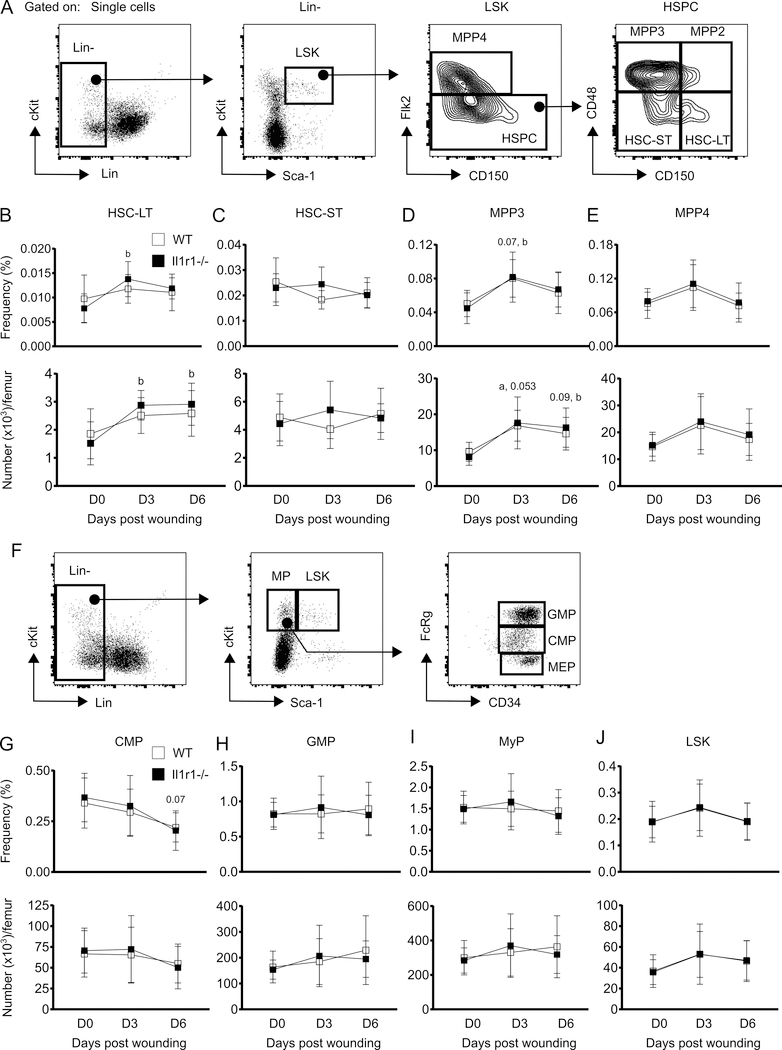
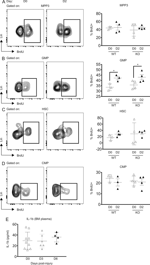
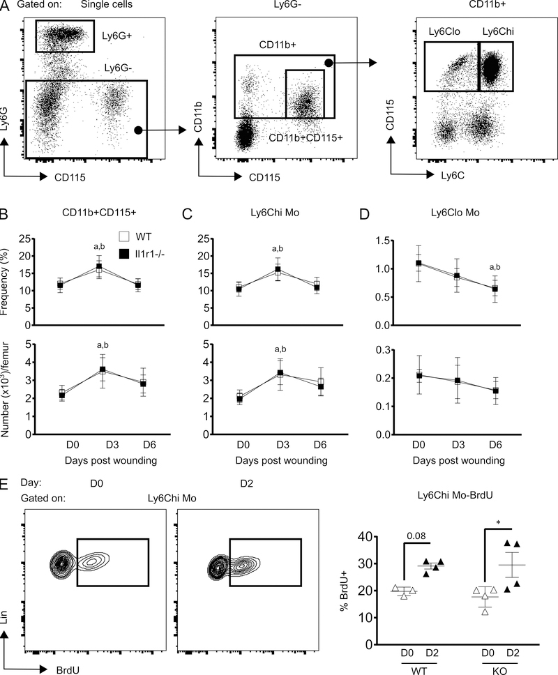
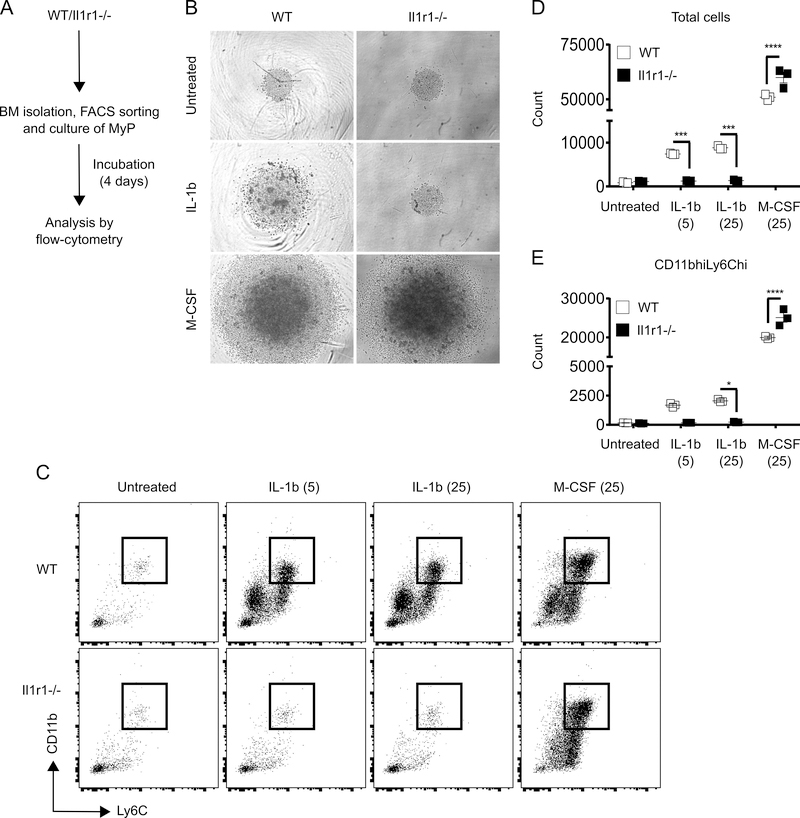
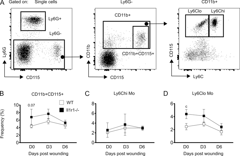
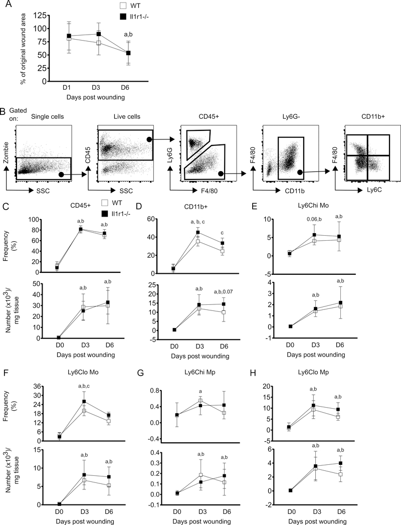
The aim of this study was to determine whether skin wounding induces monocyte (Mo) expansion in bone marrow and whether IL-1R1 signaling regulates this process. Our data show that skin wounding increases myeloid lineage-committed multipotent progenitors (MPP3 subset) and Mo in bone marrow, but this expansion is not impaired in Il1r1-/- mice. We also demonstrate that M-CSF-induced differentiation of myeloid progenitors into Mo is not impaired by the loss of IL-1R1 ex vivo, indicating that IL-R1 deficiency does not abrogate myeloid progenitor differentiation potential. In addition, we observed modestly delayed wound closure in Il1r1-/- mice associated with higher frequency of Ly6Clo Mo in the circulation at baseline and in wounds early after injury. Thus, in contrast to other models of inflammation that involve IL-1R1-dependent monopoiesis, our results demonstrate that skin wounding induces Mo progenitor and Mo expansion independently of IL-1R1 signaling.


Figures

Figure 1:
Figure 1:
Skin wounding-induced expansion of MPP3 compartment is not affected by IL-1R1 signaling deficiency. (A) Representative flow-cytograms showing gating strategy for flow-cytometry analysis of HSPC subsets in BM. (B-E) Percentage (of total BM cells (BMC); upper panels) and number (lower panels) of LT-HSC (Lin-Sca-1+cKit+(LSK)Flk2-CD48-CD150+), ST-HSC (LSK Flk2-CD48-CD150-), MPP3 (LSK Flk2-CD48+CD150-) and MPP4 (LSK Flk2+CD150-) in BM. (F) Gating strategy for MyP subsets and LSK in BM. (G-J) Percentage (of total BMC; upper panels) and number (lower panels) of CMP (Lin-Sca-1-cKit+FcRγloCD34+), GMP (Lin-Sca-1-cKit+FcRγhiCD34+), MyP (Lin-Sca-1-cKit+) and LSK in BM. Results are represented as mean ± SD, n=7 mice for each strain and each time point. a, mean value significantly different from day 0 in WT; b, mean value significantly different from day 0 in Il-1r1−/−. p≤0.05 is significant.
Figure 2:
Figure 2:
Skin wounding increases GMP proliferation IL-1R1 independently. Representative flow-cytograms showing BrdU+ cells gated on MPP3 (A), GMP (B), HSC (C) and CMP (D) (left panels) and percentage of BrdU+ cells in the corresponding cell populations (right panels). Results are represented as mean ± SD, n=3 to 4 mice for each strain and each time point. *p≤0.05. (E) IL-1β concentration in BM plasma. Results are represented as mean ± SD, n=4 to 9 mice for each time point.
Figure 3:
Figure 3:
IL-1R1 deficiency does not alter skin wounding-induced monocyte expansion in BM. (A) Representative flow-cytograms showing gating strategy for flow-cytometry analysis of Mo subsets in BM. (B-D) Percentage (of total BMC; upper panels) and number (lower panels) of total Mo (Ly6G-CD11b+CD115+), Ly6Chi Mo (Ly6G-CD11b+CD115+Ly6Chi) and Ly6Clo Mo (Ly6G-CD11b+CD115+Ly6Clo) in BM. Results are represented as mean ± SD, n=7 mice for each strain and each time point. a, mean value significantly different from day 0 in WT; b, mean value significantly different from day 0 in Il-1r1−/−. (E) Representative flow-cytograms showing BrdU+ cells gated on Ly6Chi (left panel) and percentage of Ly6Chi BrdU+ cells (right panel). Results are represented as mean ± SD, n=3 to 4 mice for each strain and each time point. *p≤0.05.
Figure 4:
Figure 4:
IL-1R1 signaling deficiency does not abrogate expansion potential of MyP. (A) Experimental scheme. (B) Representative microscopic images of MyP cultures after 4 days of incubation. Images taken at 10x magnification. (C) Representative flow-cytograms for flow-cytometry analysis of CD11bhiLy6Chi Mo after 4 days of culture. Number of total cells (D) and CD11bhiLy6Chi Mo (E) after 4 days of culture. Results are represented as mean ± SD. BM cells were collected from 2 mice for each strain and experiment was done once in triplicates. *p≤0.05, ***p≤0.001, ****p≤0.0001.
Figure 5:
Figure 5:
Frequency of Ly6Clo Mo is higher in the circulation of Il1r1−/− mice at steady state. (A) Representative flow-cytograms showing gating strategy for flow-cytometry analysis of Mo subsets in peripheral blood. (B-D) Percentage (of total BMC; upper panels) and number (lower panels) of total Mo (Ly6G-CD11b+CD115+), Ly6Chi Mo (Ly6G-CD11b+CD115+Ly6Chi) and Ly6Clo Mo (Ly6G-CD11b+CD115+Ly6Clo) in peripheral blood. Results are represented as mean ± SD, n=3 mice for each strain and each time point. c, mean value significantly different between two strains at same time point. p≤0.05 is significant.
Figure 6:
Figure 6:
IL-1R1 signaling deficiency increases Ly6Clo Mo during early wound repair. (A) Percentage of wound area as compared with day 0 wounds. ImageJ software was used to measure wound area. (B) Representative flow-cytograms showing gating strategy for flow-cytometry analysis of innate immune cells at wounds. (C-H) Percentage (of live cells; upper panels) and number (lower panels) of total leukocytes (CD45+), Mo/Mp (CD45+Ly6G-CD11b+), Ly6Chi Mo (CD45+Ly6G-CD11b+F4/80-Ly6Chi), Ly6Clo Mo (CD45+Ly6G-CD11b+F4/80-Ly6Clo), Ly6Chi Mp (CD45+Ly6G-CD11b+F4/80+Ly6Chi) and Ly6Clo Mp, (CD45+Ly6G-CD11b+F4/80+Ly6Clo) in the wounds. Results are represented as mean ± SD, n=7 mice for each strain and each time point. a, mean value significantly different from day 0 in WT; b, mean value significantly different from day 0 in Il1r1−/−; c, mean value significantly different between two strains at same time point. p≤0.05 is significant.

References

    1. Arnold L, Henry A, Poron F, Baba-Amer Y, van Rooijen N, Plonquet A, Gherardi RK, and Chazaud B 2007. Inflammatory monocytes recruited after skeletal muscle injury switch into antiinflammatory macrophages to support myogenesis. J Exp Med 204: 1057–1069. - PMC - PubMed
    1. Duffield JS, Forbes SJ, Constandinou CM, Clay S, Partolina M, Vuthoori S, Wu S, Lang R, and Iredale JP 2005. Selective depletion of macrophages reveals distinct, opposing roles during liver injury and repair. J Clin Invest 115: 56–65. - PMC - PubMed
    1. Lucas T, Waisman A, Ranjan R, Roes J, Krieg T, Muller W, Roers A, and Eming SA 2010. Differential roles of macrophages in diverse phases of skin repair. J Immunol 184: 3964–3977. - PubMed
    1. Nahrendorf M, Swirski FK, Aikawa E, Stangenberg L, Wurdinger T, Figueiredo JL, Libby P, Weissleder R, and Pittet MJ 2007. The healing myocardium sequentially mobilizes two monocyte subsets with divergent and complementary functions. J Exp Med 204: 3037–3047. - PMC - PubMed
    1. Mirza R, DiPietro LA, and Koh TJ 2009. Selective and specific macrophage ablation is detrimental to wound healing in mice. Am J Pathol 175: 2454–2462. - PMC - PubMed

Publication types

Substances