Case Reports
. 2019 May;30(5):840-853.
doi: 10.1681/ASN.2018080786. Epub 2019 Mar 25.

Mutations in NUP160 Are Implicated in Steroid-Resistant Nephrotic Syndrome

Affiliations
Case Reports

Mutations in NUP160 Are Implicated in Steroid-Resistant Nephrotic Syndrome

Feng Zhao et al. J Am Soc Nephrol. 2019 May.

Abstract

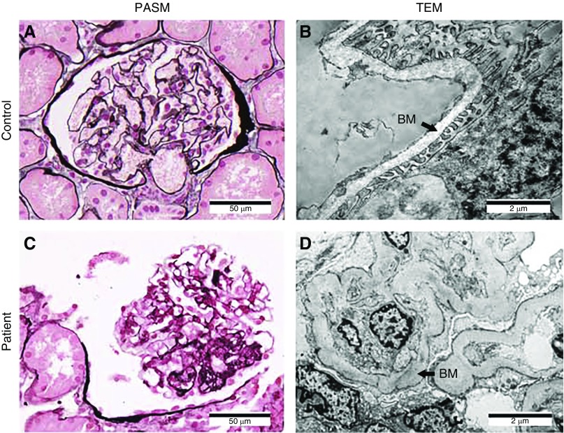
Background: Studies have identified mutations in >50 genes that can lead to monogenic steroid-resistant nephrotic syndrome (SRNS). The NUP160 gene, which encodes one of the protein components of the nuclear pore complex nucleoporin 160 kD (Nup160), is expressed in both human and mouse kidney cells. Knockdown of NUP160 impairs mouse podocytes in cell culture. Recently, siblings with SRNS and proteinuria in a nonconsanguineous family were found to carry compound-heterozygous mutations in NUP160.

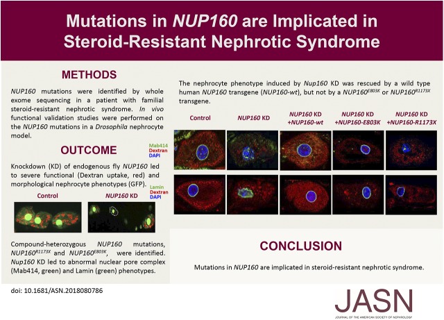
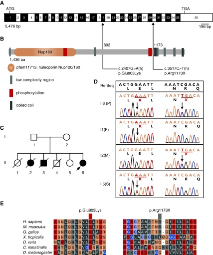
Methods: We identified NUP160 mutations by whole-exome and Sanger sequencing of genomic DNA from a young girl with familial SRNS and FSGS who did not carry mutations in other genes known to be associated with SRNS. We performed in vivo functional validation studies on the NUP160 mutations using a Drosophila model.

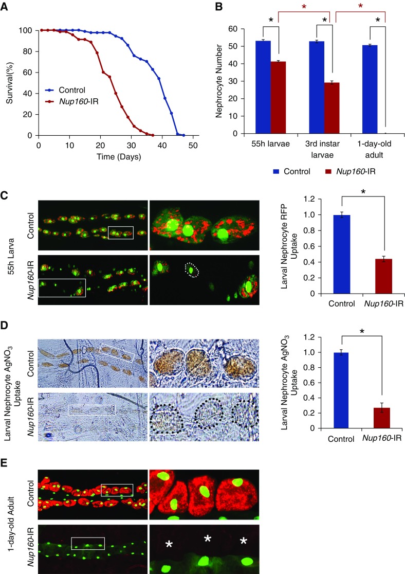
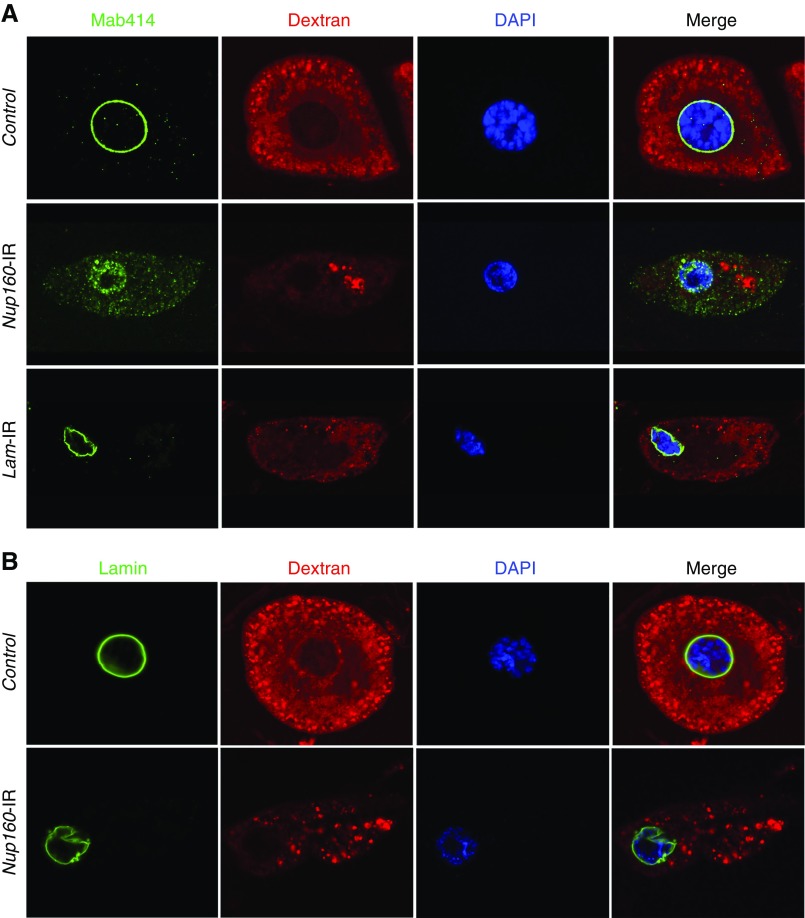
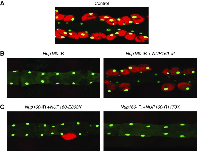
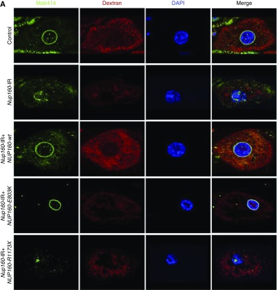
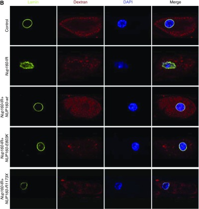
Results: We identified two compound-heterozygous NUP160 mutations, NUP160R1173× and NUP160E803K . We showed that silencing of Drosophila NUP160 specifically in nephrocytes (fly renal cells) led to functional abnormalities, reduced cell size and nuclear volume, and disorganized nuclear membrane structure. These defects were completely rescued by expression of the wild-type human NUP160 gene in nephrocytes. By contrast, expression of the NUP160 mutant allele NUP160R1173× completely failed to rescue nephrocyte phenotypes, and mutant allele NUP160E803K rescued only nuclear pore complex and nuclear lamin localization defects.

Conclusions: Mutations in NUP160 are implicated in SRNS. Our findings indicate that NUP160 should be included in the SRNS diagnostic gene panel to identify additional patients with SRNS and homozygous or compound-heterozygous NUP160 mutations and further strengthen the evidence that NUP160 mutations can cause SRNS.

Keywords: Drosophila; NUP160; genetic renal disease; human genetics; nephrocyte; nephrotic syndrome.


Figures

None
Graphical abstract
Figure 1.
Figure 1.
Compound-heterozygous mutations in NUP160, NUP160Glu803Lys and NUP160Arg1173X, were identified in an autosomal recessive family with steroid-resistant nephrotic syndrome. (A) Human NUP160 cDNA (NM_015231.2; 5476 bp) showing exons (numbered; alternating black and white), positions of start (ATG) and stop (TGA) codons, and mutation sites (arrows). (B) Human Nup160 protein (NP_056046.1; 1436 amino acids) structural domains, phosphorylation sites, and locations of missense (amino acid 803) and truncation (amino acid 1173) lesions. (C) A pedigree of the affected family (family members diagnosed with kidney disease are indicated in black). The arrow indicates the proband. (D) Compound-heterozygous missense and truncating NUP160 mutations identified from proband (II6; patient [P]) sequencing. Nucleotide and corresponding amino acid sequences are shown for wild-type (upper panel; in black) and mutant (lower panel; in green) alleles. Codons are underlined (green), and altered nucleotides and amino acids are highlighted (red). Arrows indicate positions of mutations in relation to exons and protein motifs (in A and B, respectively). Relevant sequences from surviving family members (I1, father [F]; I2, mother [M]; II5, sister [S]) are also shown. (E) Phylogenetic comparisons of amino acid sequences of Nup160 protein regions affected by the identified mutations.
Figure 2.
Figure 2.
The proband's renal tissue histology and cell ultrastructure revealed FSGS. (A and B) Control normal glomerulus tissue samples were obtained from a patient with a kidney tumor. (C and D) Patient tissues were obtained from the proband (patient II6) (Figure 1C). (A and C) Light microscopic examination of periodic acid–silver metheramine (PASM)–stained glomerulus showing focal segmental glomerular hyaline degeneration and sclerosis in the patient sample. (B and D) Transmission electron microscopy (TEM) revealed effacement of podocyte foot processes and partial thick and sclerosing glomerular basement membrane (BM; arrows) in the patient sample. Magnification, ×400 in A and C; ×12,000 in B and D.
Figure 3.
Figure 3.
Nephrocyte-specific silencing of endogenous Nup160 expression led to shortened adult fly lifespan, fewer nephrocytes, and reduced nephrocyte function. Nup160-IR flies carry a UAS-Nup160-RNAi transgene plus a nephrocyte-specific Dot-GAL4 driver that together silence endogenous fly Nup160 expression. Control flies carry only the Dot-GAL4 driver. (A) Survival curves for Nup160-IR and control adult flies (newly emerged on day 0). Nephrocyte-specific silencing of endogenous Nup160 expression was associated with reduced lifespan: Nup160-IR flies experienced 50% mortality at day 25 versus day 40 for control flies. (B) Nephrocyte-specific silencing of Nup160 induced progressive loss of nephrocytes during larval- to adult-stage development. Nephrocytes were counted in second instar (55-hour larvae) and third instar larval stages and adult flies within 24 hours postemergence. Progressively fewer nephrocytes were observed during larval-stage development, and nephrocytes were entirely absent in adult flies. For quantification, nephrocytes were counted from each of five larvae or adult flies of each genotype. The results are presented as mean±SD. *Statistical significance was defined as P<0.05. (C) Nephrocyte-specific silencing of Nup160 led to reduced uptake of fluorescently labeled marker protein. All larvae carry an MHC-ANF-RFP transgene expressing ANF-RFP marker protein in muscle cells, which is secreted into the fly hemolymph; it is normally taken up by and accumulated in nephrocytes (red fluorescence) before protein breakdown and recycling of amino acids. In addition, all flies carry a Hand-GFP transgene expressing a nuclear-localized GFP marker (green; predominantly nuclear fluorescence) in both pericardial nephrocytes (larger nuclei with faint cytoplasmic fluorescence) and cardiomyocytes (smaller nuclei). Fluorescence microscopy revealed that, in control 55-hour larvae, almost every nephrocyte took up and accumulated abundant ANF-RFP marker protein from the larval hemolymph (left panels; center panels show the boxed areas). By contrast, nephrocytes of Nup160-IR larvae expressing the Nup160-RNAi transgene showed reduced levels of ANF-RFP overall, although a minority of nephrocytes displayed essentially normal levels of RFP. Quantification of ANF-RFP uptake by control versus Nup160-IR nephrocytes is shown in right panel. Nephrocyte RFP uptake was reduced approximately 2.5-fold as a result of Nup160 gene silencing; ≥20 nephrocytes were analyzed from each of five larvae per genotype. The results are presented as mean±SD. *Statistical significance was defined as P<0.05. (D) Ingested silver nitrate (AgNO3) taken up and sequestered by third instar larval nephrocytes revealed by phase contrast microscopy. Upper left and upper center panels show abundant AgNO3 within control nephrocytes, whereas lower left and lower center panels show very reduced levels in Nup160-IR nephrocytes delineated by the dotted outline in left panels (center panels show higher-magnification views of boxed nephrocytes). Quantification of AgNO3 in control versus Nup160-IR nephrocytes is shown in right panel. Nephrocyte AgNO3 was reduced nearly fivefold as a result of Nup160 gene silencing; ≥20 nephrocytes were analyzed from each of five larvae per genotype. The results are presented as mean±SD. *Statistical significance was defined as P<0.05. (E) Nup160 gene silencing led to complete absence of adult fly nephrocytes. All flies express MHC-ANF-RFP and Hand-GFP transgenes. Fluorescence microscopy showed control adult nephrocytes with abundant cytoplasmic ANF-RFP (red) and nuclear GFP (green) as well as cardiomyocytes (smaller green nuclei; no RFP fluorescence). By contrast, no nephrocytes were observed in Nup160-IR flies expressing a UAS-Nup160-RNAi driven by Dot-Gal4, although cardiomyocytes (GFP-positive nuclei) were normal. Right panels show higher magnification views of boxed areas. *Normal locations of nephrocytes relative to cardiomyocytes.
Figure 4.
Figure 4.
Nup160 gene silencing induced nuclear pore complex (NPC) and lamin abnormalities in Drosophila nephrocytes. (A) Mab414 (green) immunolabeled components of the NPC, which in control nephrocytes, generated a sharp and highly regular ring around the nucleus (indicated by 4′,6-diamidino-2-phenylindole [DAPI] staining; blue). In nephrocytes of larvae in which Nup160 gene expression was silenced (Nup160-IR), by contrast, Mab414 immunolabeling was dispersed across the nucleus, and it was more abundant in the cytoplasm, indicating abnormal localization of NPC components. Normal ex vivo uptake of FITC-labeled 10-kD Dextran particles (red) was significantly affected by Nup160 gene silencing, indicating functional nephrocyte defects linked to abnormal NPC localization. Silencing of the Drosophila Lamin gene by nephrocyte-specific expression of a UAS-Lam-RNAi transgene driven by Dot-Gal4 (Lam-IR) led to marked abnormalities in nuclear size and shape and a severe functional deficit, but NPC localization appeared normal. (B) Mab ADL67.10 labels all isoforms of Drosophila Sf9 Lamin but does not label Lamin C. In control nephrocytes, Lamin (green) appeared as a sharp regular ring around the nucleus. Nup160 gene silencing (Nup160-IR), by contrast, did not disrupt circumferential nuclear Lamin localization (although nuclear morphology was altered as indicated by DAPI staining), but labeling was less concise and uniform than in control nephrocytes.
Figure 5.
Figure 5.
Adult nephrocyte phenotypes induced by Nup160-IR were rescued by wild type human NUP160 transgene, but not by NUP160E803K or NUP160R1173X. (A) Fluorescence micrograph shows nephrocytes of adult flies 1 day postemergence. ANF-RFP fluorescence (red) is merged with GFP (green; mostly nuclear). A GFP transgene is expressed under the control of a Hand gene enhancer (Hand-GFP) to confirm pericardial nephrocyte cell identity. All flies are transgenic for Hand-GFP. Control flies carry the Dot-Gal4 driver but no RNA interference construct. (B, left panel) Nephrocyte-specific Nup160-IR transgene expression silenced endogenous fly Nup160, leading to complete absence of adult nephrocytes. Unaffected cardiomyocytes exhibited GFP fluorescence. (B, right panel) Nephrocyte-specific expression of a wild-type human NUP160 transgene (NUP160-wt) rescued the adult nephrocyte phenotype induced by Nup160-IR. (C, left panel) Nephrocyte-specific expression of a mutant E803K NUP160 transgene (NUP160-E803K) failed to rescue the adult nephrocyte phenotype induced by Nup160-IR. (C, right panel) Nephrocyte-specific expression of a mutant R1173× NUP160 transgene (NUP160-R1173×) failed to rescue the adult nephrocyte phenotype induced by Nup160-IR.
Figure 6.
Figure 6.
Third instar larval nephrocyte phenotypes induced by Nup160-IR were rescued by wild type human NUP160, but not by NUP160E803K or NUP160R1173X. (A) Mab414 (green) immunolabeled components of the nuclear pore complex (NPC), fluorescent Dextran uptake indicated nephrocyte function (Figure 4), and 4′,6-diamidino-2-phenylindole (DAPI) staining revealed nuclear position and overall morphology. (B) Mab ADL67.10 labels all isoforms of Drosophila Sf9 Lamin (green) but does not label Lamin C. Nephrocyte-specific expression of a wild-type human NUP160 transgene (NUP160-wt) rescued all larval nephrocyte phenotypes. An NUP160E803K mutant transgene (NUP160-E803K) rescued NPC localization and Lamin morphology but not Dextran uptake. A NUP160R1173× mutant transgene (NUP160-R1173×) failed to rescue any larval nephrocyte phenotypes.
Figure 6.
Figure 6.
Third instar larval nephrocyte phenotypes induced by Nup160-IR were rescued by wild type human NUP160, but not by NUP160E803K or NUP160R1173X. (A) Mab414 (green) immunolabeled components of the nuclear pore complex (NPC), fluorescent Dextran uptake indicated nephrocyte function (Figure 4), and 4′,6-diamidino-2-phenylindole (DAPI) staining revealed nuclear position and overall morphology. (B) Mab ADL67.10 labels all isoforms of Drosophila Sf9 Lamin (green) but does not label Lamin C. Nephrocyte-specific expression of a wild-type human NUP160 transgene (NUP160-wt) rescued all larval nephrocyte phenotypes. An NUP160E803K mutant transgene (NUP160-E803K) rescued NPC localization and Lamin morphology but not Dextran uptake. A NUP160R1173× mutant transgene (NUP160-R1173×) failed to rescue any larval nephrocyte phenotypes.

Comment in

References

    1. Wiggins RC: The spectrum of podocytopathies: A unifying view of glomerular diseases. Kidney Int 71: 1205–1214, 2007 - PubMed
    1. Trautmann A, Bodria M, Ozaltin F, Gheisari A, Melk A, Azocar M, et al. . PodoNet Consortium : Spectrum of steroid-resistant and congenital nephrotic syndrome in children: The PodoNet registry cohort. Clin J Am Soc Nephrol 10: 592–600, 2015 - PMC - PubMed
    1. Preston R, Stuart HM, Lennon R: Genetic testing in steroid-resistant nephrotic syndrome: Why, who, when and how? Pediatr Nephrol 34: 195–210, 2019 - PMC - PubMed
    1. Kestilä M, Lenkkeri U, Männikkö M, Lamerdin J, McCready P, Putaala H, et al. .: Positionally cloned gene for a novel glomerular protein--nephrin--is mutated in congenital nephrotic syndrome. Mol Cell 1: 575–582, 1998 - PubMed
    1. Boute N, Gribouval O, Roselli S, Benessy F, Lee H, Fuchshuber A, et al. .: NPHS2, encoding the glomerular protein podocin, is mutated in autosomal recessive steroid-resistant nephrotic syndrome. Nat Genet 24: 349–354, 2000 - PubMed

Publication types

LinkOut - more resources