. 2019 Mar 25;9(1):5102.
doi: 10.1038/s41598-019-41060-0.

The cochaperone CHIP marks Hsp70- and Hsp90-bound substrates for degradation through a very flexible mechanism

Affiliations

The cochaperone CHIP marks Hsp70- and Hsp90-bound substrates for degradation through a very flexible mechanism

Lucía Quintana-Gallardo et al. Sci Rep. .

Abstract

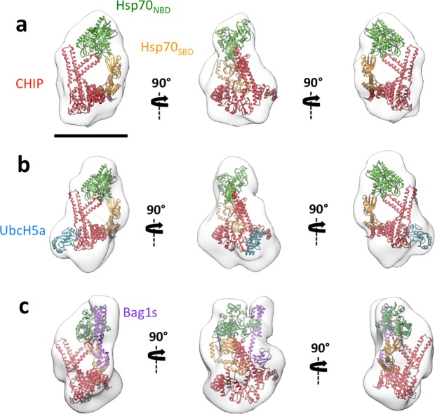
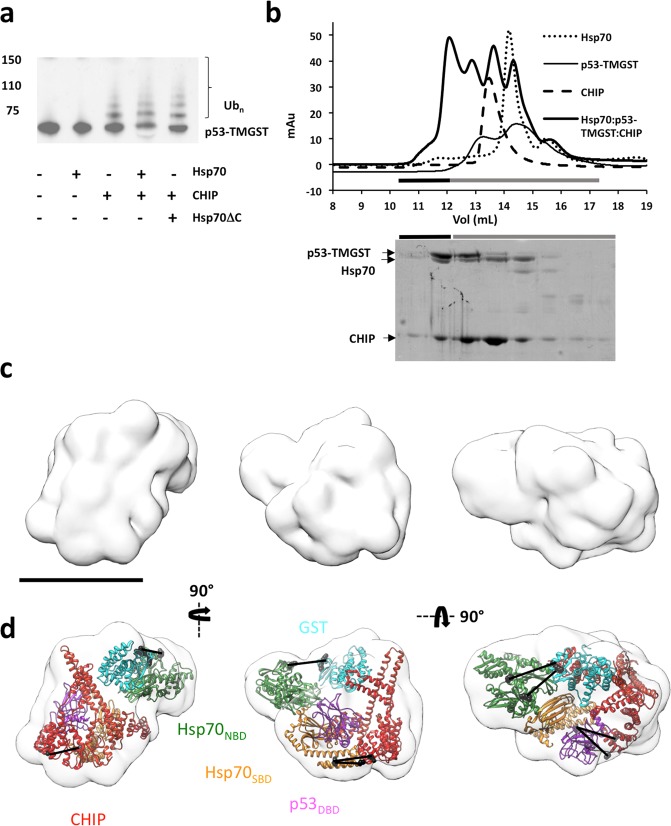
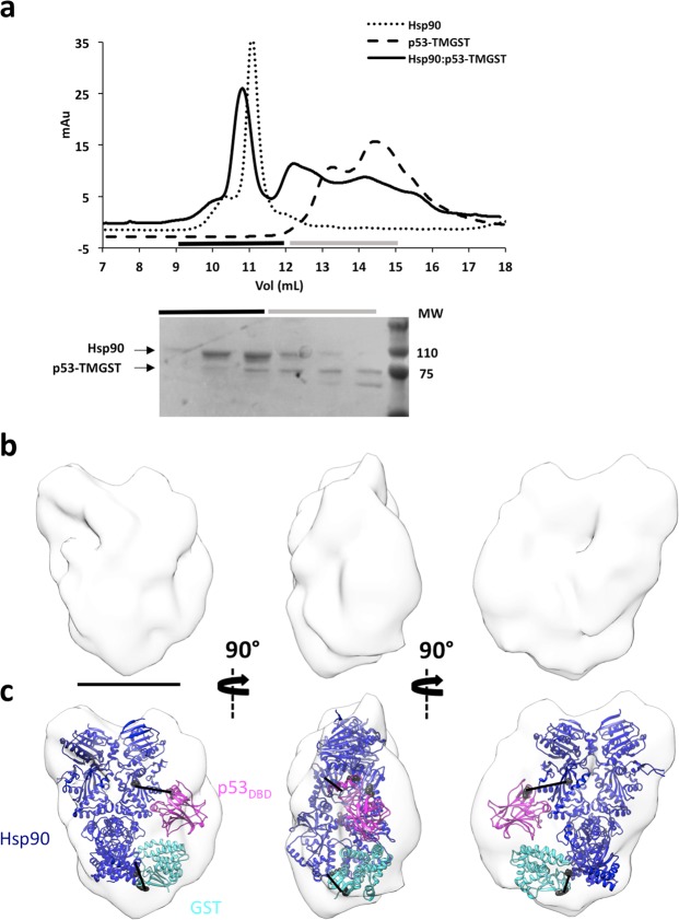
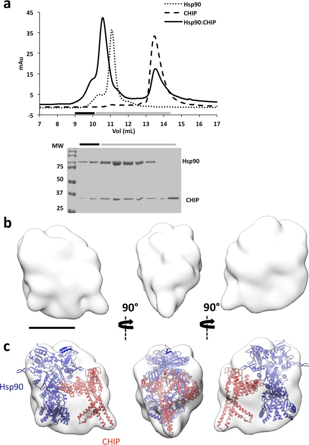
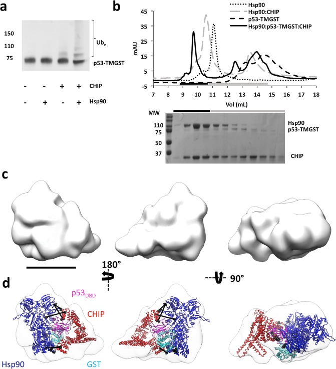
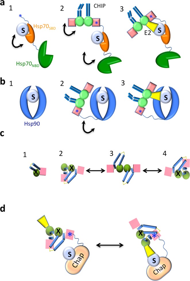
Some molecular chaperones are involved not only in assisting the folding of proteins but also, given appropriate conditions, in their degradation. This is the case for Hsp70 and Hsp90 which, in concert with the cochaperone CHIP, direct their bound substrate to degradation through ubiquitination. We generated complexes between the chaperones (Hsp70 or Hsp90), the cochaperone CHIP and, as substrate, a p53 variant containing the GST protein (p53-TMGST). Both ternary complexes (Hsp70:p53-TMGST:CHIP and Hsp90:p53-TMGST:CHIP) ubiquitinated the substrate at a higher efficiency than in the absence of the chaperones. The 3D structures of the two complexes, obtained using a combination of cryoelectron microscopy and crosslinking mass spectrometry, showed the substrate located between the chaperone and the cochaperone, suggesting a ubiquitination mechanism in which the chaperone-bound substrate is presented to CHIP. These complexes are inherently flexible, which is important for the ubiquitination process.


Conflict of interest statement

The authors declare no competing interests.

Figures

Figure 1
Figure 1
Isolation and structural characterisation of the Hsp70:p53-TMGST complex. (a) Top, size-exclusion profile of the complex (continuous line) compared with the profiles for Hsp70 (dotted line) and p53-TMGST (broken line). Bottom, the selected fractions were analysed by SDS-PAGE and stained with Coomassie blue. (b) Three orthogonal views of the Hsp70:p53-TMGST complex 3D reconstruction. (c) The same views with docking of the chaperone Hsp70 (Hsp70NBD (pdb 5aqz; green) and Hsp70 SBD (pdb 4po2; orange)) and the substrate p53-TMGST (p53DBD (pdb 2ocj; pink) and GST (pdb 1m99; cian)). The DTSSP crosslinks in the images are described in Supplementary Fig. 2c. Only crosslinks between residues located in regions with known atomic structure are depicted in the figure. Bar, 100 Å for (b,c).
Figure 2
Figure 2
3D reconstructions of Hsp70:CHIP-based complexes. The same three orthogonal views of (a) 3D reconstruction of the Hsp70:CHIP complex, with docking of Hsp70 (Hsp70NBD, green and Hsp70SBD, orange) and CHIP (pdb 2c2l; red), (b) 3D reconstruction of the Hsp70:CHIP:UbcH5a complex, with docking of Hsp70, CHIP and UbCH5a (pdb 2oxq; dark blue), (c) 3D reconstruction of the Hsp70:CHIP:Bag1s complex, with docking of Hsp70 and Bag1s (from the atomic structure of the Hsp70NBD:Bag1 complex, pdb: 4hwi; pink), and CHIP. Bar, 100 Å.
Figure 3
Figure 3
Isolation, ubiquitination activity, and structural characterisation of the Hsp70:p53-TMGST:CHIP complex. (a) p53-TMGST ubiquitination assay mediated by Hsp70 and CHIP. For the western blot, an anti-p53 antibody was used. (b) Formation of the Hsp70:p53-TMGST:CHIP complex. Top, size-exclusion profile of the complex (thick continuous line) compared with the profiles for Hsp70 (dotted line), p53-TMGST (thin continuous line), and CHIP (broken line). Bottom, the selected fractions were analysed by SDS-PAGE and stained with Coomassie Blue. (c) Three orthogonal views of the Hsp70:p53-TMGST:CHIP complex 3D reconstruction. (d) The same views with docking of Hsp70 (Hsp70NBD and Hsp70 SBD), p53-TMGST (p53DBD and GST), and CHIP. The DTSSP crosslinks in the images are described in Supplementary Fig. 5c. Only crosslinks between residues located in regions with known atomic structure are depicted in the figure. Colours as in Fig. 2. Bar, 100 Å for (c,d).
Figure 4
Figure 4
Isolation and structural characterisation of the Hsp90:p53-TMGST complex. (a) Top, size-exclusion profile of the complex (continuous line) compared with the profiles for Hsp90 (dotted line) and p53-TMGST (broken line). Bottom, the selected fractions were analysed by SDS-PAGE and stained with Coomassie Blue. (b) Three orthogonal views of the Hsp90:p53-TMGST complex 3D reconstruction. (c) The same views with docking of Hsp90 (pdb 5fwk; dark blue) and p53-TMGST. The DTSSP crosslinks in the images are described in Supplementary Fig. 6c. Only crosslinks between residues located in regions with known atomic structure are depicted in the figure. Bar, 100 Å for (b,c).
Figure 5
Figure 5
Isolation and structural characterisation of the Hsp90:CHIP complex. (a) Top, size-exclusion profile of the complex (continuous line) compared with the profiles for Hsp90 (dotted line) and p53-TMGST (broken line). Bottom, the selected fractions were analysed by SDS-PAGE and stained with Coomassie Blue. (b) Three orthogonal views of the Hsp90:p53-TMGST complex 3D reconstruction. (c) The same views, with docking of Hsp90 and CHIP. Bar, 100 Å for (b,c).
Figure 6
Figure 6
Isolation, ubiquitination activity and structural characterisation of the Hsp90:p53-TMGST:CHIP complex. (a) p53-TMGST ubiquitination assay mediated by Hsp90 and CHIP. For the western blot, an anti-p53 antibody was used. (b) Formation of the Hsp90:p53-TMGST:CHIP complex. Top, size-exclusion profile of the complex (continuous line) compared with the profiles for Hsp90 (dotted line), p53-TMGST (broken lines), and the Hsp90:CHIP complex (grey broken line). Bottom, the selected fractions were analysed by SDS-PAGE and stained with Coomassie Blue. (c) Three orthogonal views of the Hsp90:p53-TMGST:CHIP complex 3D reconstruction. (d) The same views with docking of Hsp90, p53-TMGST (p53DBD and GST), and CHIP. The DTSSP and DSSO crosslinks depicted in the images are described in Supplementary Fig. 7c. Only crosslinks between residues located in regions with known atomic structure are depicted in the figure. Not all the crosslinks are depicted in every view. Bar, 100 Å for (c,d).
Figure 7
Figure 7
p53-TMGST ubiquitination mediated by chaperones Hsp70, Hsp90, and their variants with shorter, disordered C-terminal sequences. (a) Top, sequence of the full-length C-terminal region of Hsp70 and its shorter variant. Bottom, western blot of a ubiquitination experiment using an anti-p53 antibody. (b) Top, sequence of the full-length C-terminal region of Hsp90 and its shorter variant. Bottom, western blot of a ubiquitination experiment using an anti-p53 antibody.
Figure 8
Figure 8
Flexibility in the interaction between chaperone, substrate and CHIP. (a) For Hsp70: (1) a flexible complex is formed through weak interactions between the Hsp70SBD and the substrate (S) (black arrows represent flexibility). (2) CHIP binds to Hsp70:S through the Hsp70 IEEVD motif (blue star) at the C-terminus of the chaperone and the TPR domain of one of the CHIP monomers. The EEVD motif is at the end of a long unstructured region (32 residues), which makes the Hsp70-CHIP interaction very flexible. (3) CHIP movements, together with binding of an E2 ubiquitin-conjugating enzyme (i.e., UbCH5a; yellow), allow substrate ubiquitination. (b) For Hsp90: (1) a flexible complex is formed through weak interactions between the Hsp90 substrate-binding region and the substrate. (2) CHIP binds to Hsp90:S through the MEEVD motif at the end of the long, unstructured region (32 residues) of one of the Hsp90 monomers and the TPR domain of one of the CHIP monomers, generating a flexible ternary complex with the substrate positioned between Hsp90 and CHIP. (3) CHIP movements, together with binding of an E2 enzyme, allow substrate ubiquitination. (c) Model of the conformational changes undergone by CHIP (structure observed from the top). The crystal structure of full-length CHIP is that of an asymmetric dimer. (1) Each monomer is composed of a TPR domain (pink cube), a U-box domain (green sphere), and a dimerization domain (blue cylinders). Molecular dynamics studies have shown that the monomer in solution is stabilized through TPR-Ubox interactions which block the E2 binding site (X). (2) In the dimer, the two helix-coils come together such that one bends and generates two smaller, kinked helices. The dimer switches between the two possible asymmetric conformations (2 and 4) through a short-lived symmetric conformation (3). Dimer formation activates CHIP by unlocking one of the two E2 binding sites (the one without X). (d) Model of chaperone-bound, substrate ubiquitination mediated by CHIP and an E2 enzyme. When interacting with the chaperone:substrate complex, CHIP switches between the two asymmetric conformations described in (c), and E2 binding can alternate between the two potential E2-binding sites. Whereas the conformation on the left is that found in the 3D-reconstructed CHIP-based complexes in this study, the only productive interaction is that on the right, as it permits contact between the E2 enzyme and the substrate.

References

    1. Hipp MS, Park S-H, Hartl FU. Proteostasis impairment in protein-misfolding and -aggregation diseases. Trends Cell Biol. 2014;24:506–514. doi: 10.1016/j.tcb.2014.05.003. - DOI - PubMed
    1. Arndt V, Rogon C, Höhfeld J. To be, or not to be - Molecular chaperones in protein degradation. Cell. Mol. Life Sci. 2007;64:2525–2541. doi: 10.1007/s00018-007-7188-6. - DOI - PMC - PubMed
    1. Hartl FU, Bracher A, Hayer-Hartl M. Molecular chaperones in protein folding and proteostasis. Nature. 2011;475:324–332. doi: 10.1038/nature10317. - DOI - PubMed
    1. Stankiewicz M, Nikolay R, Rybin V, Mayer MP. CHIP participates in protein triage decisions by preferentially ubiquitinating Hsp70-bound substrates. FEBS J. 2010;277:3353–3367. doi: 10.1111/j.1742-4658.2010.07737.x. - DOI - PubMed
    1. McDonough H, Patterson C. CHIP: a link between the chaperone and proteasome systems. Cell Stress Chaperones. 2003;8:303–308. doi: 10.1379/1466-1268(2003)008<0303:CALBTC>2.0.CO;2. - DOI - PMC - PubMed

Publication types

Substances