. 2019 Mar 25;9(1):5052.
doi: 10.1038/s41598-019-41513-6.

Gene Duplication Associated with Increased Fluconazole Tolerance in Candida auris cells of Advanced Generational Age

Affiliations

Gene Duplication Associated with Increased Fluconazole Tolerance in Candida auris cells of Advanced Generational Age

Somanon Bhattacharya et al. Sci Rep. .

Abstract

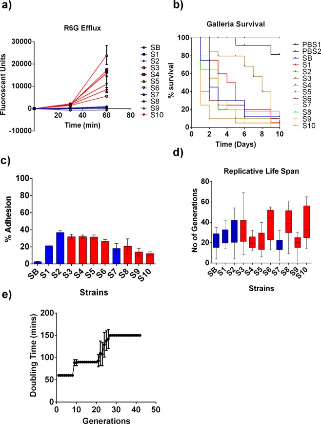
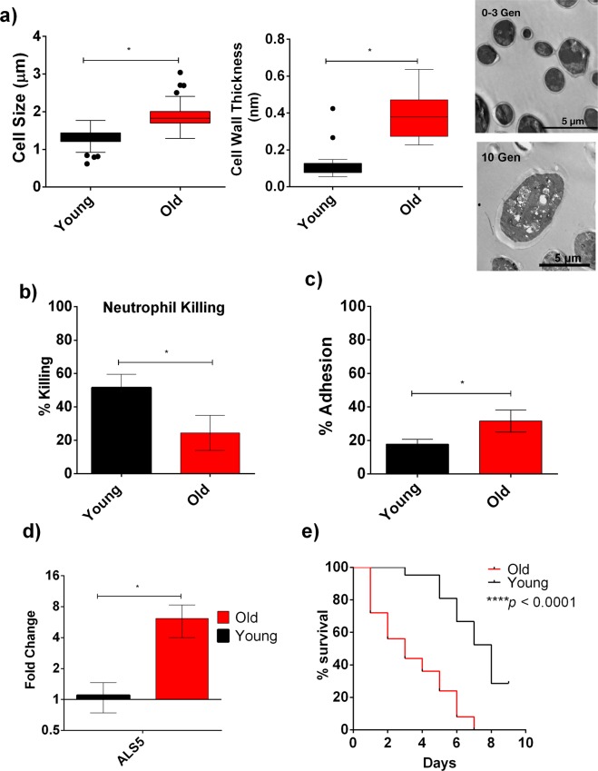
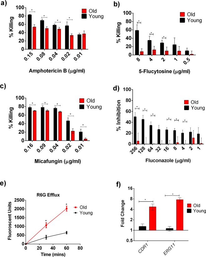
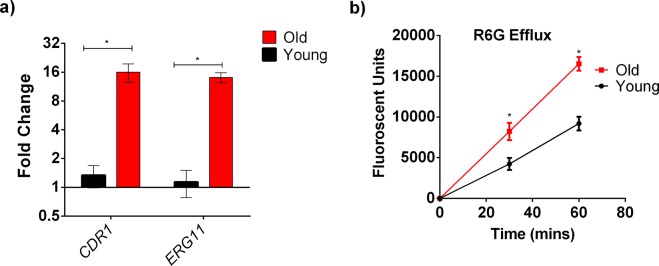
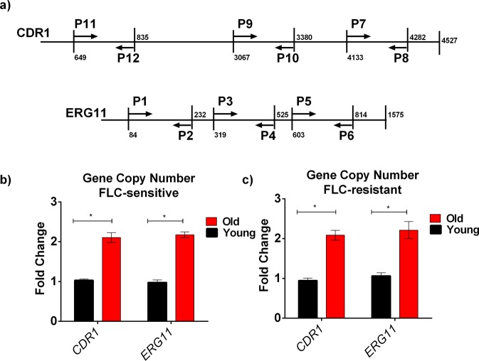
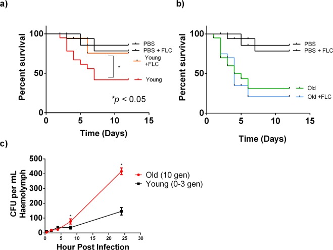
Candida auris is an emerging multi-drug resistant yeast that causes systemic infections. Here we show that C. auris undergoes replicative aging (RA) that results from asymmetric cell division and causes phenotypic differences between mother and daughter cells similar to other pathogenic yeasts. Importantly, older C. auris cells (10 generations) exhibited higher tolerance to fluconazole (FLC), micafungin, 5- flucytosine and amphotericin B compared to younger (0-3 generation) cells. Increased FLC tolerance was associated with increased Rhodamine 6G (R6G) efflux and therapeutic failure of FLC in a Galleria infection model. The higher efflux in the older cells correlated with overexpression of the efflux pump encoding gene CDR1 (4-fold). In addition, 8-fold upregulation of the azole target encoding gene ERG11 was noted in the older cells. Analysis of genomic DNA from older cells by qPCR indicates that transient gene duplication of CDR1 and ERG11 causes the observed age-dependent enhanced FLC tolerance in C. auris strains. Furthermore, older cells exhibited a thickened cell wall, decreased neutrophil killing (24% vs 50%), increased epithelial cell adhesion (31.6% vs 17.8%) and upregulation of adhesin protein Als5p. Thus, this study demonstrates that transient gene duplication can occur during RA, causing increased FLC tolerance in old C. auris cells.


Conflict of interest statement

The authors declare no competing interests.

Figures

Figure 1
Figure 1
Antifungal Resistance Mechanism and Virulence Phenotypes Observed in the C. auris Clinical Isolates: (a) R6G assay was performed on all the 11 C. auris strains. Red lines signify FLC resistant isolates, blue lines signify FLC susceptible isolates. R6G assay was done in triplicate, and error bars signify standard deviation. (b) C. auris isolates show variable virulence in Galleria. 105 cells from each isolate were injected into 20 larvae (n = 20) and their survival was observed every 24 h for 10 days. Two sets of 20 larvae (labelled in the figure as PBS1 and PBS2) were injected with only 1X PBS as controls. (c) Percent adherence of all the 11 C. auris strains to HeLa cells was measured. Red and blue bars signify FLC resistant and susceptible strains respectively. The assay was done in triplicate and error bars signify standard deviation. (d) Representative data of RLS of C. auris clinical isolates. RLS for each strain was determined for n = 20 cells. The minimum and maximum values of all data are indicated by the ends of the whiskers of box plots. The middle box is the interquartile range divided at the median. The Red and blue bars signify FLC resistant and susceptible strains respectively. (e) Change in doubling time of strain S1 was observed with increase in generational age (n = 20). Error bars represent Standard deviation and One-Way Anova was done to determine the significance. Doubling times significantly changed between Gen 1 and Gen 10 cells, Gen 1 and Gen 20, and Gen 1 and Gen 30 (*p < 0.05).
Figure 2
Figure 2
Increased Virulence of 10 Gen Old C. auris Cells from FLC- Susceptible S1: (a) Cell size and Cell wall thickness of the old (10 Gen) cells were significantly greater compared to young (0–3 Gen) cells. Cell wall thickness (n = 23) and Cell Size (n = 150) were measured by ImageJ software from the microscopic images of individual young and old cells. Student’s t-test was performed to analyze the significance (*p < 0.001). Transmission Electron Microscopic (TEM) image of the old (10 Gen) cells of C. auris showing increased cell wall thickness compared to the young (0–3 Gen) cells. (b) Percent Neutrophil-mediated Killing of young (0–3 Gen: black bar) and old (10 Gen: red bar) cells of C. auris. The assay was done in triplicate and error bars signify standard deviation. Student’s t-test with Welch’s correction was performed to determine the significance; *p = 0.0266. (c) Percent adhesion of young (0–3 Gen: black bar) and old (10 Gen: red bar) cells of C. auris. The assay was done in triplicate and error bars signify standard deviation. Student’s t-test with Welch’s correction was performed to determine the significance; *p = 0.0291. (d) qPCR analysis to study the expression of ALS5 in young and old cells. ACT1 gene was used as an internal control and the data was normalized to gene expression in young cells. The assay was done in triplicate and error bars signify standard deviation. Multiple t-test was performed using Holm-Sidak method to analyze the significance; *p = 0.016. (e) Virulence of young (0–3 Gen) and old (10 Gen) cells of C. auris in Galleria. 20 larvae were infected separately with 105 young and old cells. Log rank test was used to determine the statistical significance.
Figure 3
Figure 3
Increased Antifungal Tolerance of 10 Gen Old C. auris cells from FLC- Susceptible S1 strain: Old (10 Gen) cells are significantly more tolerant to antifungals (a) AMB, (b) 5FC, (c) MF, (d) FLC. Black bars signify young cells, while red bars signify old cells. The assays were done in triplicate and error bars signify standard deviation. Multiple t-Test was performed using Holm-Sidak method to analyze the significance; *p < 0.05; (e) R6G efflux assay performed on young (black line) and old (red line) in triplicate and error bars signify standard deviation. Student’s t-test with Welch’s correction was performed to determine the significance; *p < 0.05. (f) qPCR analysis to study the expression of CDR1 and ERG11 in young and old cells. ACT1 gene was used as an internal control. The data was normalized to the gene expression in young cells. The assay was done in triplicate and error bars signify standard deviation. Multiple t-test was performed using Holm-Sidak method to analyze the significance; *p < 0.05.
Figure 4
Figure 4
Increased Antifungal Tolerance of 10 Generation Old C. auris cells from FLC- Resistant S9 strain: (a) qPCR analysis to study the expression of CDR1 and ERG11 in young and old cells. ACT1 gene was used as an internal control. The data was normalized to the gene expression in young cells. The assay was done in biological triplicate and error bars signify standard deviation. Multiple t-test was performed using Holm-Sidak method to analyze the significance; *p < 0.05. (b) R6G efflux assay performed on young (black line) and old (red line) in triplicate and error bars signify standard deviation. Student’s t-test with Welch’s correction was performed to determine the significance; *p < 0.05.
Figure 5
Figure 5
qPCR Analysis to Study the Increase in Copy Numbers of CDR1 and ERG11 in Old C. auris cells isolated from strain S1.: (a) Cartoon showing the location of the oligonucleotides used in this analysis. The primers span 3 different regions of each genes. “P” denotes primer. All primers listed in Table S1. Genomic DNA was isolated from both old (10 generation) cells and young (0–3 generation) cells of FLC-sensitive strain S1 (b) and FLC-resistant strain S9 (c). Data shows the gene copy numbers of CDR1 and ERG11 using oligo sets P7-P8 (CDR1) and P5-P6 (ERG11) ACT1 was used as a control for the experiment and the data was normalized to the gene copy number of young cells. The assay was performed in triplicate and error bars signify standard deviation. Multiple t-test was performed using Holm-Sidak method to analyze the significance; *p < 0.05.
Figure 6
Figure 6
FLC Treatment of Galleria infected with Young (0–3 Gen) and Old (10 Gen) cells of C. auris: 20 larvae were infected separately with 105 (a) young and (b) old cells. 1 µg/ml of FLC was injected to the larvae infected with young and old population on the same day of infection. Log rank test was used to determine the statistical significance. (c) Cells from old (10 Gen) S1 strain were significantly retained more in the haemolymph (*p < 0.05) when compared to young cells at different time points. Haemolymph was collected from 15 larvae over the period of infections (0, 0.5, 2, 4, 8 and 24 h) as described and plated in YPD plates. CFUs were counted and plotted after 48 h of incubation at 37 °C. For each time point, 5 × 104 young and old cells from isolate S1 was infected into 15 larvae. Error bars represent standard deviation in the CFU counts. (n = 15) for each infection time point. Multiple t-test was performed using Holm-Sidak method to determine the statistical significance (*p < 0.05).

References

    1. Chowdhary A, Sharma C, Meis JF. Candida auris: A rapidly emerging cause of hospital-acquired multidrug-resistant fungal infections globally. PLoS Pathogens. 2017;13:e1006290. doi: 10.1371/journal.ppat.1006290. - DOI - PMC - PubMed
    1. Ben-Ami, R. et al. Multidrug-Resistant Candida haemulonii and C. auris, Tel Aviv, Israel. Emerging Infectious Diseases23, 10.3201/eid2302.161486 (2017). - PMC - PubMed
    1. Sears D, Schwartz BS. Candida auris: An emerging multidrug-resistant pathogen. International Journal of Infectious Diseases: IJID: official publication of the International Society for Infectious Diseases. 2017;63:95–98. doi: 10.1016/j.ijid.2017.08.017. - DOI - PubMed
    1. Kathuria S, et al. Multidrug-Resistant Candida auris Misidentified as Candida haemulonii: Characterization by Matrix-Assisted Laser Desorption Ionization-Time of Flight Mass Spectrometry and DNA Sequencing and Its Antifungal Susceptibility Profile Variability by Vitek 2, CLSI Broth Microdilution, and Etest Method. Journal of Clinical Microbiology. 2015;53:1823–1830. doi: 10.1128/JCM.00367-15. - DOI - PMC - PubMed
    1. Arendrup MC, Patterson TF. Multidrug-Resistant Candida: Epidemiology, Molecular Mechanisms, and Treatment. The Journal of Infectious Diseases. 2017;216:S445–S451. doi: 10.1093/infdis/jix131. - DOI - PubMed

Publication types

LinkOut - more resources