. 2019 Mar 22:4:13.
doi: 10.1038/s41541-019-0107-7. eCollection 2019.

A prophylactic α-Gal-based glycovaccine effectively protects against murine acute Chagas disease

Affiliations

A prophylactic α-Gal-based glycovaccine effectively protects against murine acute Chagas disease

Susana Portillo et al. NPJ Vaccines. .

Abstract

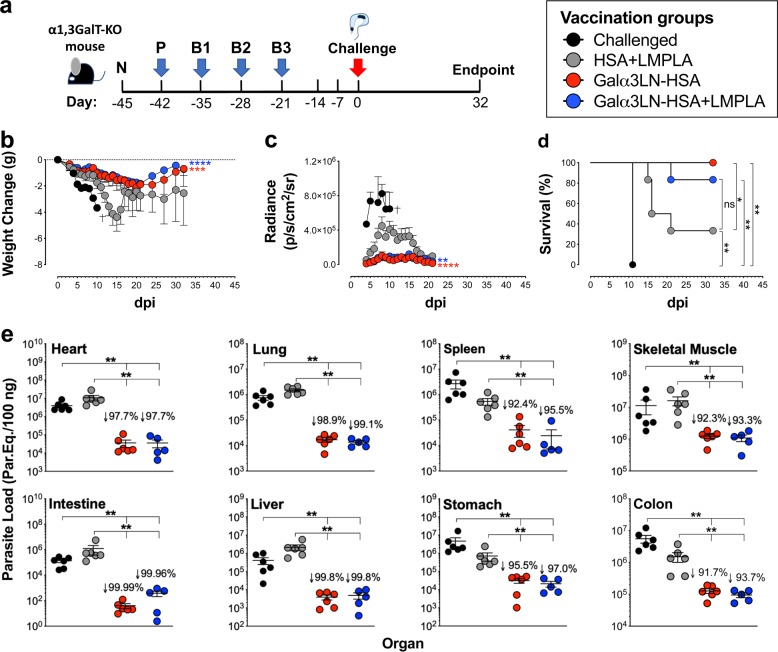
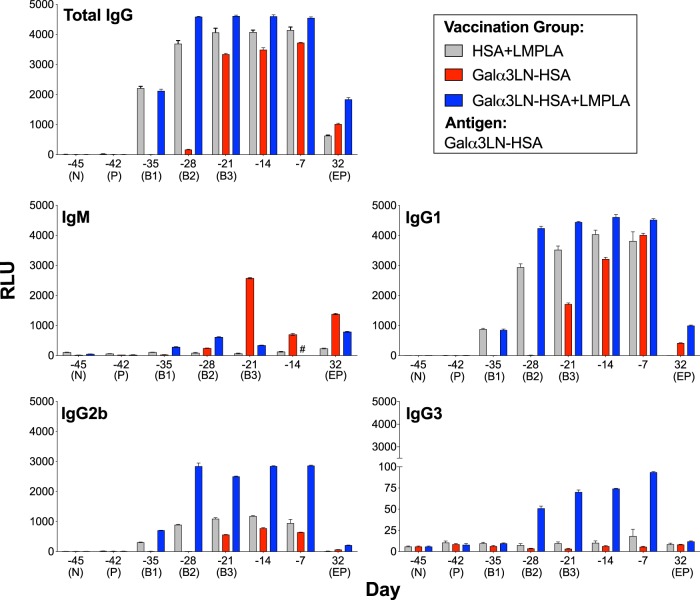
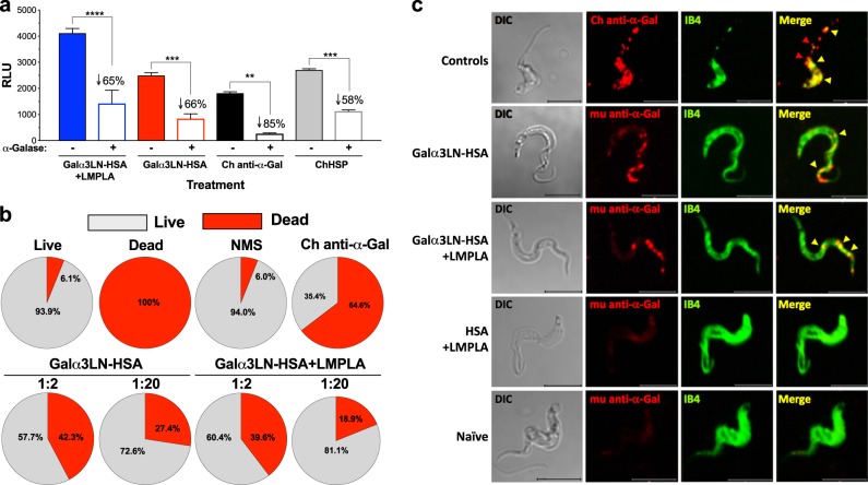
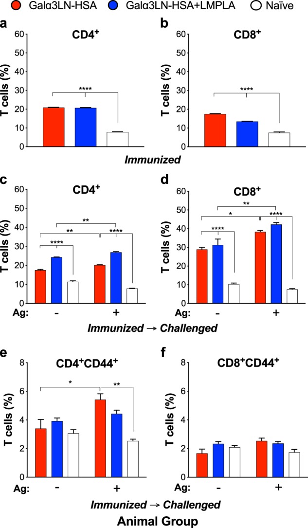
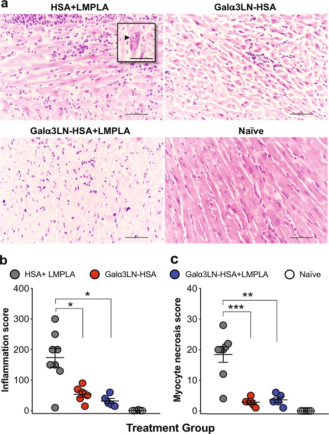
Chagas disease (ChD), caused by the hemoflagellate parasite Trypanosoma cruzi, affects six to seven million people in Latin America. Lately, it has become an emerging public health concern in nonendemic regions such as North America and Europe. There is no prophylactic or therapeutic vaccine as yet, and current chemotherapy is rather toxic and has limited efficacy in the chronic phase of the disease. The parasite surface is heavily coated by glycoproteins such as glycosylphosphatidylinositol (GPI)-anchored mucins (tGPI-mucins), which display highly immunogenic terminal nonreducing α-galactopyranosyl (α-Gal)-containing glycotopes that are entirely absent in humans. The immunodominant tGPI-mucin α-Gal glycotope, the trisaccharide Galα1,3Galβ1,4GlcNAc (Galα3LN), elicits high levels of protective T. cruzi-specific anti-α-Gal antibodies in ChD patients in both the acute and chronic phases. Although glycoconjugates are the major parasite glycocalyx antigens, they remain completely unexplored as potential ChD vaccine candidates. Here we investigate the efficacy of the T. cruzi immunodominant glycotope Galα3LN, covalently linked to a carrier protein (human serum albumin (HSA)), as a prophylactic vaccine candidate in the acute model of ChD, using the α1,3-galactosyltransferase-knockout (α1,3GalT-KO) mouse, which mimics the human immunoresponse to α-Gal glycotopes. Animals vaccinated with Galα3LN-HSA were fully protected against lethal T. cruzi challenge by inducing a strong anti-α-Gal antibody-mediated humoral response. Furthermore, Galα3LN-HSA-vaccinated α1,3GalT-KO mice exhibited significant reduction (91.7-99.9%) in parasite load in all tissues analyzed, cardiac inflammation, myocyte necrosis, and T cell infiltration. This is a proof-of-concept study to demonstrate the efficacy of a prophylactic α-Gal-based glycovaccine for experimental acute Chagas disease.


Conflict of interest statement

The authors declare no competing interests.

Figures

Fig. 1
Fig. 1
α1,3GalT-WT and α1,3GalT-KO mice exhibit different immune responses to T. cruzi infection. a Parasitemia in α1,3GalT-WT and α1,3GalT-KO mice infected (intraperitoneally (i.p.)) with 1 × 103 tissue culture-derived trypomastigotes (TCTs) (Y strain). b Kaplan–Meier survival rate curve of animals infected with 1 × 104 TCTs (Y strain). c Anti-α-Gal Ab levels as measured by chemiluminescent enzyme-linked immunosorbent assay using Galα3LN-BSA as antigen. RLU relative luminescence units. Infected C57BL/6 α1,3GalT-WT mice were compared to knockout (KO) mice by two-way analysis of variance (ANOVA) with Tukey’s multiple comparison test. *p < 0.05; ****p < 0.0001. d Serum cytokine profile of α1,3GalT-WT and α1,3GalT-KO mice infected (i.p.) with 1 × 103 TCTs and followed up for 28 days. N naive. Lysis of TCTs (1 × 107/mL) by serum e or purified anti-α-Gal Abs f from α1,3GalT-WT and α1,3GalT-KO mice infected (i.p.) with 1 × 103 TCTs. Unbound immunoglobulin G (IgG), non-anti-α-Gal IgG antibodies (Abs) (flow through) from Synsorb 115 immunoaffinity chromatography; anti-α-Gal IgG, anti-α-Gal Abs eluted from Synsorb 115. Inhibition of parasite host cell invasion g and intracellular amastigote proliferation h. LLC-MK2 cells were infected with Y strain TCTs (multiplicity of infection = 10), for 2 h at 37 °C, in the presence or not of 100 μg/well of murine non-anti-α-Gal IgG Abs (Unbound IgG from Synsorb 115) or purified murine anti-α-Gal IgG Abs or control (None, medium alone). Number of infected cells per 1000 cells was evaluated after staining with 4,6-diamidino-2-phenylindole. a, b, f, g One-way ANOVA with Dunn’s multiple comparisons test. ***p < 0.001; ****p < 0.0001. The p values in a, b pertain to the mean values of the α1,3GalT-KO group in comparison with the α1,3GalT-WT throughout the course of the experiment. Error bars indicate S.E.M
Fig. 2
Fig. 2
Vaccination with Galα3LN-HSA or Galα3LN-HSA+LMPLA protects α1,3GalT-KO mice against T. cruzi infection. a Experimental design scheme. N naive (before prime); P prime; B1–B3, boosts 1–3. Three weeks after B3, immunized α1,3GalT-KO mice were challenged (intraperitoneally) with 1 × 105T. cruzi CL Brener-luc tissue culture-derived trypomastigotes. Experimental endpoint was 32 days postinfection. b Weight change in grams (g) assessed in immunized (Galα3LN-HSA+/−LMPLA) and control (HSA+LMPLA) α1,3GalT-KO mice daily after parasite challenge. One-way analysis of variance (ANOVA) with Dunn’s multiple comparisons test was performed comparing the immunized (Galα3LN-HSA+/−LMPLA) groups with the control group (HSA+LMPLA); p values indicate the significance for the whole group. *p < 0.05; **p < 0.01; ***p < 0.001; ****p < 0.0001. Death of all animals (n = 3) in the challenged-only group is indicated (†). Error bars indicate S.E.M. c In vivo bioluminescence imaging of α1,3GalT-KO mice was performed to quantify parasite burden in the whole body following parasite challenge. Images were obtained daily and radiance (p/s/cm2/sr) was plotted. One-way ANOVA with Dunn’s multiple comparisons test was performed comparing the immunized (Galα3LN-HSA+/−LMPLA) groups with the control group (HSA+LMPLA); p values indicate the significance for the whole group. **p < 0.01; ****p < 0.0001. Error bars indicate S.E.M. Death of all animals (n = 3) in the challenged-only group is indicated (†). d Kaplan–Meier survival rate was assessed and recorded in each animal group (n = 6 per group in all groups, except for group “Challenged”) after parasite challenge. Log-rank (Mantel–Cox) test was performed to compare the immunized groups (Galα3LN-HSA+/−LMPLA) with the control groups (Challenged-only and HSA-LMPLA). *p < 0.05; **p < 0.01. e Determination of parasite load by quantitative real-time PCR in the heart, lung, spleen, skeletal muscle, intestine, liver, stomach, and colon, using satellite DNA of T. cruzi. Parasite load is expressed as parasite equivalents per 100 ng of tissue. Reduction of parasite load was calculated by dividing the mean of experimental vaccines by the HSA+LMPLA control group. Error bars indicate S.E.M. of triplicate determinations. Student’s t test with Mann–Whitney was performed to compare the immunized groups (Galα3LN-HSA+/−LMPLA) with the control groups (Challenged-only and HSA-LMPLA). **p < 0.01. The p values in b, c pertain to the mean values of each immunized group in comparison with the HSA+LMPLA control group throughout the course of the experiment
Fig. 3
Fig. 3
Kinetics of humoral immune response elicited by Galα3LN-HSA, Galα3LN-HSA+LMPLA, and HSA+LMPLA. Chemiluminescent enzyme-linked immunosorbent assay (CL-ELISA) reactivity of mouse sera from α1,3GalT-KO mice injected with Galα3LN-HSA, Galα3LN-HSA+LMPLA, or HSA+LMPLA using Galα3LN-HSA as immobilized antigen. For immunoglobulin M (IgM) measurement, prior to the CL-ELISA we depleted IgG antibodies by preincubating each serum sample with a 15-μL bead suspension mixture of protein A-Sepharose 4B and protein G-Sepharose, both fast flow (A:G, 1:1, v/v), and recombinant human serum albumin (HSA; 1 mg/mL), for 1 h at 37 °C on rotatory shaker. Error bars indicate S.E.M. of triplicate determinations. Sample no longer available is indicated (#). RLU relative luminescence units
Fig. 4
Fig. 4
Anti-Galα3LN-HSA Abs are α-Gal-specific, have lytic activity, and recognize parasite surface glycotopes. a Chemiluminescent enzyme-linked immunosorbent assay reactivity of sera from α1,3GalT-KO mice vaccinated with Galα3LN-HSA+/−LMPLA before and after green coffee bean α-galactosidase (α-Galase) treatment of the immobilized Galα3LN-HSA antigen. Purified human Chagas (Ch) anti-α-Gal immunoglobulin G (IgG) antibodies (Abs) and human serum pool from chronic Ch disease patients (ChHSP) were used as controls. RLU relative luminescence units. One-way ANOVA with Sidak’s multiple comparison test was performed comparing groups before (−) and after (+) α-galactosidase (α-Galase) treatment; p values indicate the significance of group before and after treatment. **p < 0.01; ***p < 0.001; ****p < 0.0001. b Percentage of lysis of tissue culture-derived trypomastigotes (TCTs) incubated with sera from α1,3GalT-KO mice vaccinated with Galα3LN-HSA+/−LMPLA. c Confocal microscopy using murine (mu) anti-α-Gal IgG Abs purified from mice vaccinated with Galα3LN-HSA+/−LMPLA. Bandeiraea simplicifolia Isolectin IB4 was used to label all terminal α-Gal residues on TCT surface. Human Ch anti-α-Gal IgG Abs were used as positive control. Bar 5 μm
Fig. 5
Fig. 5
Vaccination with Galα3LN-HSA+/−LMPLA induce T cell activation. Splenocytes were collected from mice immunized with Galα3LN-HSA or Galα3LN-HSA+LMPLA and immunized-challenged mice, followed by ex vivo stimulation with 20 μg/mL Galα3LN-HSA. a, b Percentage of antigen-specific CD4+ and CD8+ T cells, respectively, in immunized mice. c, d Percentage of CD4+ and CD8+ T cells, respectively, in immunized-challenged mice. e, f Percentage of memory CD4+CD44+ and CD8+CD44+ T cells, respectively, in immunized-challenged mice. One-way analysis of variance (ANOVA) compared with naive group was obtained for the immunized groups; Two-way ANOVA compared with the naive group was obtained for the immunized-challenged groups; *p < 0.05; **p < 0.01; ****p < 0.0001. Error bars indicate S.E.M. of triplicate determinations
Fig. 6
Fig. 6
Histopathology analysis. a Micrographs of heart sections harvested at the endpoint and stained with hematoxylin and eosin. Inset, arrowhead indicates an amastigote nest. b Myocardial inflammatory and mononuclear cell infiltrates were manually scored. c Myocyte necrosis was manually counted in 25 fields (×400). Magnification ×40, bar 100 μm. Statistical significance was calculated by unpaired two-tailed Mann–Whitney test, comparing Galα3LN-HSA+/−LMPLA group to HSA+LMPLA group. p values indicate the significance *p < 0.05; **p < 0.01; ***p < 0.001

References

    1. Bern C. Chagas' disease. N. Engl. J. Med. 2015;373:456–466. doi: 10.1056/NEJMra1410150. - DOI - PubMed
    1. Rassi A, Jr, Rassi A, Marin-Neto JA. Chagas disease. Lancet. 2010;375:1388–1402. doi: 10.1016/S0140-6736(10)60061-X. - DOI - PubMed
    1. Urbina JA. Recent clinical trials for the etiological treatment of chronic chagas disease: advances, challenges and perspectives. J. Eukaryot. Microbiol. 2015;62:149–156. doi: 10.1111/jeu.12184. - DOI - PubMed
    1. Ribeiro I, et al. New, improved treatments for Chagas disease: from the R&D pipeline to the patients. PLoS Negl. Trop. Dis. 2009;3:e484. doi: 10.1371/journal.pntd.0000484. - DOI - PMC - PubMed
    1. Beaumier CM, et al. Status of vaccine research and development of vaccines for Chagas disease. Vaccine. 2016;34:2996–3000. doi: 10.1016/j.vaccine.2016.03.074. - DOI - PubMed

LinkOut - more resources