

Functional vascular anatomy of the peritoneum in health and disease
- PMID: 30911618
- PMCID: PMC6328070
- DOI: 10.1515/pp-2016-0015
Functional vascular anatomy of the peritoneum in health and disease
Abstract
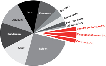
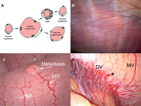
The peritoneum consists of a layer of mesothelial cells on a connective tissue base which is perfused with circulatory and lymphatic vessels. Total effective blood flow to the human peritoneum is estimated between 60 and 100 mL/min, representing 1-2 % of the cardiac outflow. The parietal peritoneum accounts for about 30 % of the peritoneal surface (anterior abdominal wall 4 %) and is vascularized from the circumflex, iliac, lumbar, intercostal, and epigastric arteries, giving rise to a quadrangular network of large, parallel blood vessels and their perpendicular offshoots. Parietal vessels drain into the inferior vena cava. The visceral peritoneum accounts for 70 % of the peritoneal surface and derives its blood supply from the three major arteries that supply the splanchnic organs, celiac and superior and inferior mesenteric. These vessels give rise to smaller arteries that anastomose extensively. The visceral peritoneum drains into the portal vein. Drugs absorbed are subject to first-pass hepatic metabolism. Peritoneal inflammation and cancer invasion induce neoangiogenesis, leading to the development of an important microvascular network. Anatomy of neovessels is abnormal and characterized by large size, varying diameter, convolution and blood extravasation. Neovessels have a defective ultrastructure: formation of large "mother vessels" requires degradation of venular and capillary basement membranes. Mother vessels give birth to numerous "daughter vessels". Diffuse neoangiogenesis can be observed before appearance of macroscopic peritoneal metastasis. Multiplication of the peritoneal capillary surface by neoangiogenesis surface increases the part of cardiac outflow directed to the peritoneum.
Keywords: anatomy; peritoneum; vascular anatomy; vessel.
Figures
References
-
- Verger C. Peritoneal ultrastructure. In: Nolph KD, editor. Peritoneal dialysis. Boston, MA: Martinus Nijhoff, 1985:95–113.
-
- Henderson LW. The problem of peritoneal membrane area and permeability. Kidney Int 1973;3:409–10. - PubMed
-
- Mion CM, Boen ST. Analysis of factors responsible for the formation of adhesions during chronic peritoneal dialysis. Am J Med Sci 1965;250:675–9. - PubMed
-
- Knapowski J, Feder E, Simon M, Zabel M. Evaluation of the participation of parietal peritoneum in dialysis: physiological morphological and pharmacological data. Proc Eur Dial Trans Assoc 1979;16:155–64. - PubMed
Publication types
LinkOut - more resources
Full Text Sources