. 2019 Mar 15:2:105.
doi: 10.1038/s42003-019-0343-4. eCollection 2019.

Mutations in the PKM2 exon-10 region are associated with reduced allostery and increased nuclear translocation

Affiliations

Mutations in the PKM2 exon-10 region are associated with reduced allostery and increased nuclear translocation

Tsan-Jan Chen et al. Commun Biol. .

Abstract

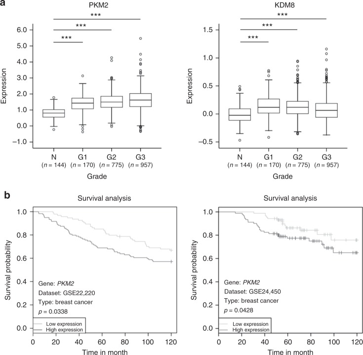
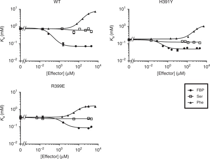
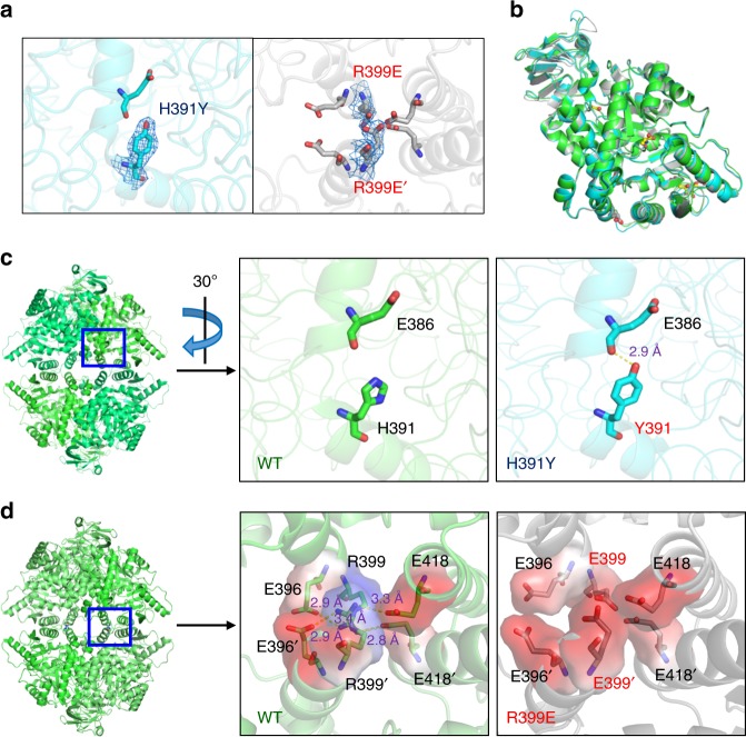
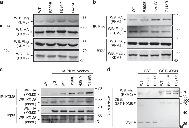
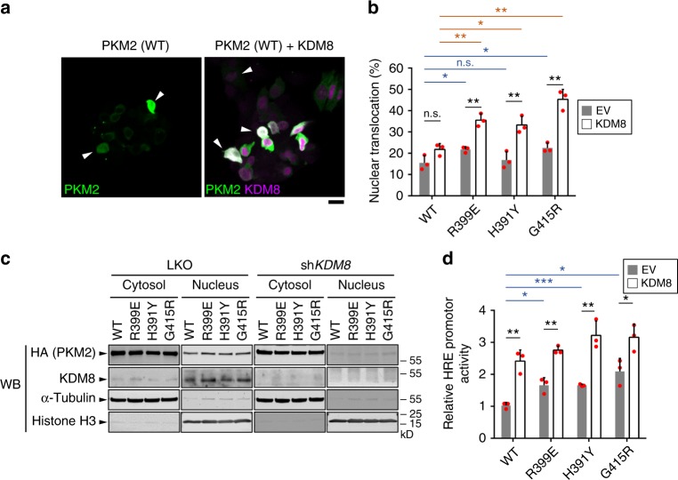
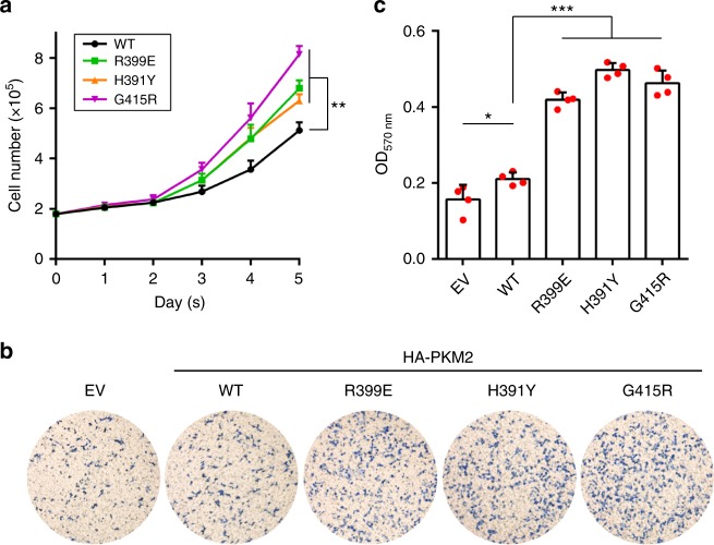
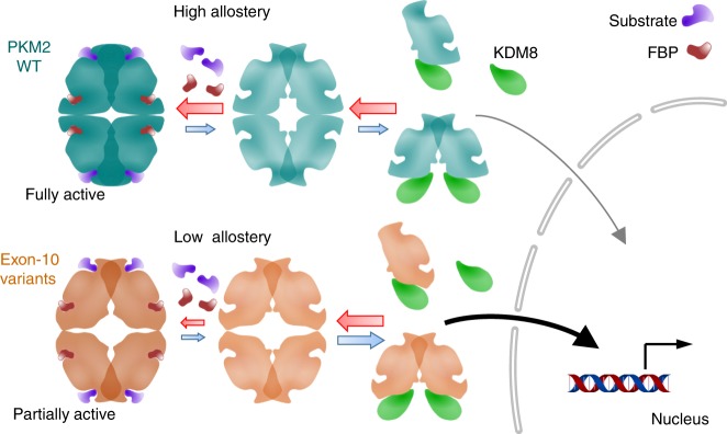
PKM2 is a key metabolic enzyme central to glucose metabolism and energy expenditure. Multiple stimuli regulate PKM2's activity through allosteric modulation and post-translational modifications. Furthermore, PKM2 can partner with KDM8, an oncogenic demethylase and enter the nucleus to serve as a HIF1α co-activator. Yet, the mechanistic basis of the exon-10 region in allosteric regulation and nuclear translocation remains unclear. Here, we determined the crystal structures and kinetic coupling constants of exon-10 tumor-related mutants (H391Y and R399E), showing altered structural plasticity and reduced allostery. Immunoprecipitation analysis revealed increased interaction with KDM8 for H391Y, R399E, and G415R. We also found a higher degree of HIF1α-mediated transactivation activity, particularly in the presence of KDM8. Furthermore, overexpression of PKM2 mutants significantly elevated cell growth and migration. Together, PKM2 exon-10 mutations lead to structure-allostery alterations and increased nuclear functions mediated by KDM8 in breast cancer cells. Targeting the PKM2-KDM8 complex may provide a potential therapeutic intervention.


Conflict of interest statement

The authors declare no competing interests.

Figures

Fig. 1
Fig. 1
Clinical relevance of PKM2 and KDM8 in cancers. a PKM2 and KDM8 were overexpressed using METABRIC (the Molecular Taxonomy of Breast Cancer International Consortium) database (***p < 0.0001, two-tailed t-test). The source data is available in Supplementary Data 1. b Negative survival outcomes in two data sets from Table 1. N, normal. G1 to G3, grade 1 to grade 3
Fig. 2
Fig. 2
The coupling effect between the PEP binding affinity and an allosteric effector by the linked-function analysis. The profile of Ka values for PEP of PKM2 (wild-type, H391Y, or R399E) vs. an effector (FBP, Ser, or Phe) was measured. Data from three independent experiments are presented as mean ± SD. WT, wild-type
Fig. 3
Fig. 3
Structural analysis of PKM2 wild-type and variants. a The 2Fo-Fc electron density map of H391Y and R399E. The map is contoured at the 1.1-σ level. b Superposition between wild-type (R-state, green), H391Y (cyan), and R399E (gray) monomers. Mutated residues are shown as heavy sticks. FBP, serine, and oxalate are drawn as ball-and-stick models. The carbon, oxygen, and nitrogen atoms are colored yellow, red, and blue, respectively. c Structural analysis of wild-type PKM2 and H391Y shows that H391Y possesses a strong hydrogen bond contact between Y391 and E386. The right panel is a zoomed region of 30 degree rotation clockwise of the left panel. d Comparison between wild-type PKM2 and R399E shows that R399E fails to form a salt-bridge network among R399, E396, and E418 at the C−C interface. The right panel is a zoomed region of the left panel. Residues (E386, H391, Y391, R399, E399, E396, and E418) are shown as sticks. The oxygen and nitrogen atoms are colored red and blue, respectively. wild-type (PDB: 3SRD; green); H391Y (PDB: 4YJ5, this study; cyan); and R399E (PDB: 5X0I, this study; gray)
Fig. 4
Fig. 4
PKM2 exon-10 variants show increased interaction with KDM8. a, b Co-immunoprecipitation (IP) assay was conducted by co-transfecting MCF7 cells with Flag-KDM8 plus a HA-tagged PKM2 (HA-PKM2) protein as indicated, followed by IP with anti-HA (a) or anti-Flag (b) and Western blotting analysis. c IP assay was conducted by transfecting MCF7 cells with a HA-PKM2 as indicated, followed by IP with anti-KDM8 and the subsequent Western blotting analysis. d GST pull-down assays were performed with GST or GST-KDM8 and a His-tagged PKM2 (His-PKM2) protein as indicated. WB, Western blotting analysis; EV, empty vector; endo., endogenous; CBB, Coomassie brilliant blue; WT, wild-type
Fig. 5
Fig. 5
KDM8 promotes the nuclear translocation and transactivation activity of allostery-insensitive PKM2 variants. a, b MCF7 cells were transfected with HA-PKM2s (wild-type, R399E, H391Y, and G415R) or co-transfected with HA-PKM2 plus Flag-KDM8, followed by staining with anti-HA (HA-PKM2, green) and anti-Flag (Flag-KDM8, magenta). Representative images of the nuclear translocation of PKM2 in wild-type-PKM2-expressing cells in the absence or presence of KDM8 are shown in a. Arrowheads indicate the cells with detectable nuclear localization of PKM2. Bar 20 μm. The mean percentage of nuclear-localized PKM2 cells over PKM2-expressing cells from three preparations (n ≥ 50) for each PKM2 type (wild-type, R399E, H391Y, and G415R) was determined as shown in b (wild-type vs. R399E, p = 0.043; wild-type vs. H391Y, p = 0.040; wild-type + KDM8 vs. H391Y + KDM8, p = 0.015). c MCF7 cells were transfected with HA-PKM2s with or without knocking down endogenous KDM8 as indicated. Cells were then fractionated followed by Western blotting analysis. Histone H3 and α-tubulin were used as the marker for nucleus and cytosol, respectively. d Nuclear transactivation activity (wild-type vs. R399E, p = 0.013; wild-type vs. H391Y, p = 0.011; G415R vs. G415R + KDM8, p = 0.034). Statistical significance was evaluated using the two-tailed t-test. *p< 0.05; **p< 0.01; ***p< 0.001; n.s., p > 0.05; LKO, pLKO shRNA control. Bar plots are shown in mean ± SD. WT, wild-type
Fig. 6
Fig. 6
Overexpression of PKM2 exon-10 variants promotes cell proliferation and migration. a, b MCF7 cells were transfected with each HA-PKM2 mutant as indicated, followed by cell number counting over a five-day period (a) and migration assay (b). Representative images of migration are shown (b). c Quantitation of cell migration activity from b (EV vs. wild-type, p = 0.042). Statistical significance was evaluated using the t-test (two-tailed). *p< 0.05; **p< 0.01; ***p< 0.001. Plots are shown in mean ± SD. WT, wild-type
Fig. 7
Fig. 7
The proposed model that depicts the mechanistic basis of PKM2 exon-10 mutations in allosteric regulation and KDM8-mediated nuclear translocation. WT, wild-type

References

    1. Mazurek S. Pyruvate kinase type M2: a key regulator of the metabolic budget system in tumor cells. Int. J. Biochem. Cell Biol. 2011;43:969–980. doi: 10.1016/j.biocel.2010.02.005. - DOI - PubMed
    1. Iqbal MA, Gupta V, Gopinath P, Mazurek S, Bamezai RN. Pyruvate kinase M2 and cancer: an updated assessment. FEBS Lett. 2014;588:2685–2692. doi: 10.1016/j.febslet.2014.04.011. - DOI - PubMed
    1. Dayton TL, Jacks T, Vander Heiden MG. PKM2, cancer metabolism, and the road ahead. EMBO Rep. 2016;17:1721–1730. doi: 10.15252/embr.201643300. - DOI - PMC - PubMed
    1. Garnett ME, Dyson RD, Dost FN. Pyruvate kinase isozyme changes in parenchymal cells of regenerating rat liver. J. Biol. Chem. 1974;249:5222–5226. - PubMed
    1. Mazurek S, Boschek CB, Hugo F, Eigenbrodt E. Pyruvate kinase type M2 and its role in tumor growth and spreading. Semin. Cancer Biol. 2005;15:300–308. doi: 10.1016/j.semcancer.2005.04.009. - DOI - PubMed

Publication types

MeSH terms