. 2019 Jun;291(3):689-697.
doi: 10.1148/radiol.2019182095. Epub 2019 Mar 26.

Cost-effectiveness of Intraoperative MRI for Treatment of High-Grade Gliomas

Affiliations

Cost-effectiveness of Intraoperative MRI for Treatment of High-Grade Gliomas

Peter Abraham et al. Radiology. 2019 Jun.

Abstract

Background Intraoperative MRI has been shown to improve gross-total resection of high-grade glioma. However, to the knowledge of the authors, the cost-effectiveness of intraoperative MRI has not been established. Purpose To construct a clinical decision analysis model for assessing intraoperative MRI in the treatment of high-grade glioma. Materials and Methods An integrated five-state microsimulation model was constructed to follow patients with high-grade glioma. One-hundred-thousand patients treated with intraoperative MRI were compared with 100 000 patients who were treated without intraoperative MRI from initial resection and debulking until death (median age at initial resection, 55 years). After the operation and treatment of complications, patients existed in one of three health states: progression-free survival (PFS), progressive disease, or dead. Patients with recurrence were offered up to two repeated resections. PFS, valuation of health states (utility values), probabilities, and costs were obtained from randomized controlled trials whenever possible. Otherwise, national databases, registries, and nonrandomized trials were used. Uncertainty in model inputs was assessed by using deterministic and probabilistic sensitivity analyses. A health care perspective was used for this analysis. A willingness-to-pay threshold of $100 000 per quality-adjusted life year (QALY) gained was used to determine cost efficacy. Results Intraoperative MRI yielded an incremental benefit of 0.18 QALYs (1.34 QALYs with intraoperative MRI vs 1.16 QALYs without) at an incremental cost of $13 447 ($176 460 with intraoperative MRI vs $163 013 without) in microsimulation modeling, resulting in an incremental cost-effectiveness ratio of $76 442 per QALY. Because of parameter distributions, probabilistic sensitivity analysis demonstrated that intraoperative MRI had a 99.5% chance of cost-effectiveness at a willingness-to-pay threshold of $100 000 per QALY. Conclusion Intraoperative MRI is likely to be a cost-effective modality in the treatment of high-grade glioma. © RSNA, 2019 Online supplemental material is available for this article. See also the editorial by Bettmann in this issue.


Figures

Figure 1:
Figure 1:
Mobile intraoperative MRI suite that allows surgeons to perform examination during surgery without moving the patient. This unit is suspended by ceiling tracks, allowing movement in two separate surgical suites. Image courtesy of University of California San Diego Health.
Figure 2:
Figure 2:
Microsimulation model flowchart. Simulated patients were initially randomized to resection with or without intraoperative MRI. Initial resection may be complicated by intraoperative hemorrhage, which could require return to the operating department. Greater extent of resection (gross-total resection > subtotal resection) indicates higher probability of progression-free survival, although all patients are at some risk of cancer progression after operation. Patients that have persistent progressive disease (ie, recurrences) are offered up to two additional resections (three total). Patient death can occur at any time throughout the simulation. HGG = high-grade glioma, iMRI = intraoperative MRI, QALY = quality-adjusted life year.
Figure 3:
Figure 3:
Overall survival for patients who did and did not undergo intraoperative MRI. Median survival for patients who underwent intraoperative MRI was 26 months compared with 22 months for patients who did not undergo intraoperative MRI. A nonuniform scale on the horizontal axis was used to more easily compare the model’s monthly survival rates with survival rates at monthly and yearly intervals in the literature.
Figure 4:
Figure 4:
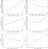
Selected one-way sensitivity analyses. Progression-free survival advantage, A, less than about 1 month and, D, overall survival advantage less than 0.5 months; B, probability of aphasia less than 9.36%, and, C, gross-total resection rate greater than 60% in the group that did not undergo intraoperative MRI made intraoperative MRI cost-ineffective at a willingness-to-pay threshold of $100 000. E, Duration of postoperative aphasia (maximum 84 months) can considerably reduce the incremental cost-effectiveness ratio (ICER; as low as $42 131 per quality-adjusted life year [QALY]), though intraoperative MRI is cost-effective regardless of the number of months of postoperative aphasia. F, Intraoperative MRI ceases to be cost-effective at a cost greater than $3500 per operation, which was about 1.5 times our upper-limit cost.
Figure 5:
Figure 5:
Cost-effectiveness acceptability curve demonstrates the result of 1000 simulated patients transitioning through the model. To determine the effect of varying multiple model parameters simultaneously on the incremental cost-effectiveness ratio, distributions in Table E1 (online) were sampled 5000 times per patient. Operation with intraoperative MRI overtakes operation without intraoperative MRI as the more cost-effective modality at an incremental cost-effectiveness ratio of $77 000 per quality-adjusted life year (QALY; before the willingness-to-pay threshold of $100 000 per QALY).

Comment in

References

    1. Johannesen TB, Langmark F, Lote K. Cause of death and long-term survival in patients with neuro-epithelial brain tumours: a population-based study. Eur J Cancer 2003;39(16):2355–2363. - PubMed
    1. van den Bent MJ, Weller M, Wen PY, Kros JM, Aldape K, Chang S. A clinical perspective on the 2016 WHO brain tumor classification and routine molecular diagnostics. Neuro Oncol 2017;19(5):614–624. - PMC - PubMed
    1. Ostrom QT, Gittleman H, Xu J, et al. CBTRUS statistical report: primary brain and other central nervous system tumors diagnosed in the United States in 2009-2013. Neuro Oncol 2016;18(suppl_5):v1–v75. - PMC - PubMed
    1. Brown TJ, Brennan MC, Li M, et al. Association of the extent of resection with survival in glioblastoma: a systematic review and meta-analysis. JAMA Oncol 2016;2(11):1460–1469. - PMC - PubMed
    1. Trifiletti DM, Alonso C, Grover S, Fadul CE, Sheehan JP, Showalter TN. Prognostic implications of extent of resection in glioblastoma: analysis from a large database. World Neurosurg 2017;103:330–340. - PubMed

Publication types

MeSH terms