. 2019 Apr 23;13(4):4278-4289.
doi: 10.1021/acsnano.8b09403. Epub 2019 Apr 4.

Anti-Biofilm Activity of Graphene Quantum Dots via Self-Assembly with Bacterial Amyloid Proteins

Affiliations

Anti-Biofilm Activity of Graphene Quantum Dots via Self-Assembly with Bacterial Amyloid Proteins

Yichun Wang et al. ACS Nano. .

Abstract

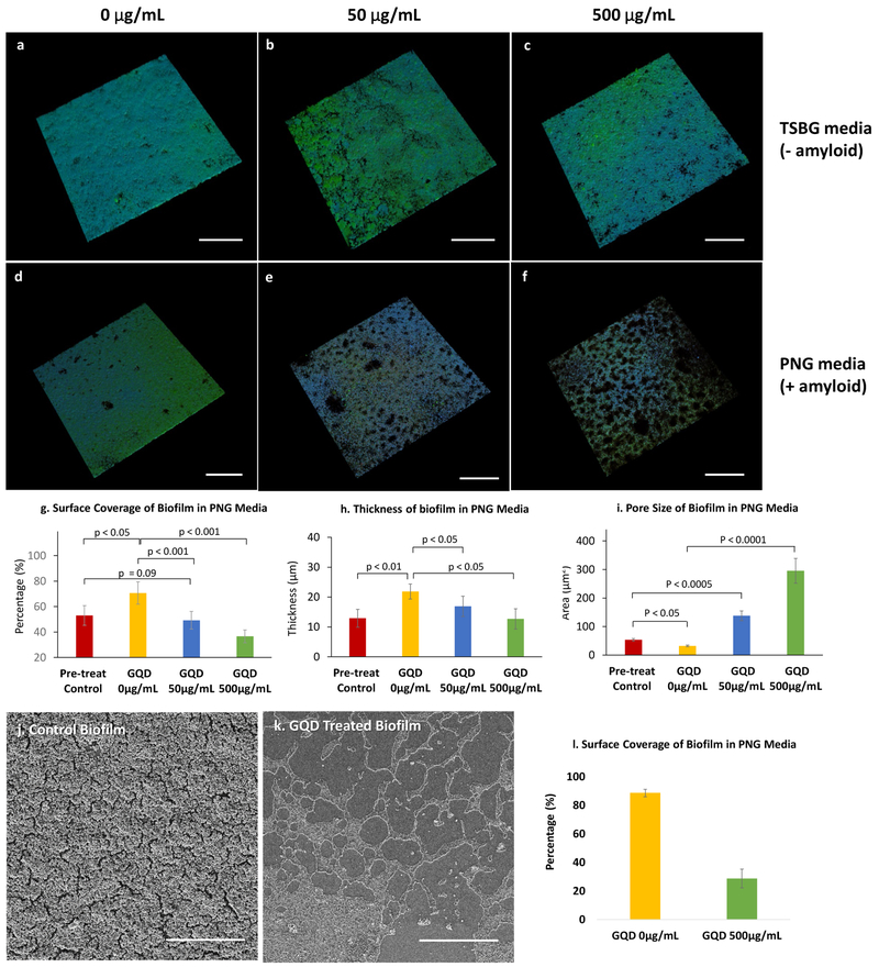
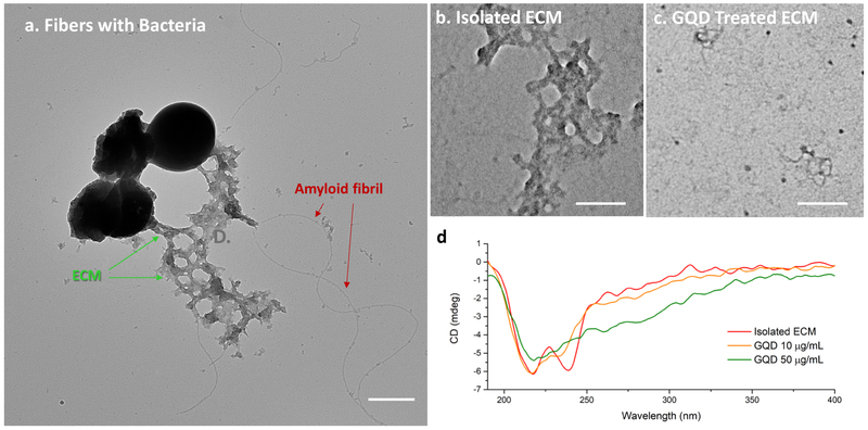
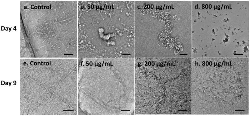
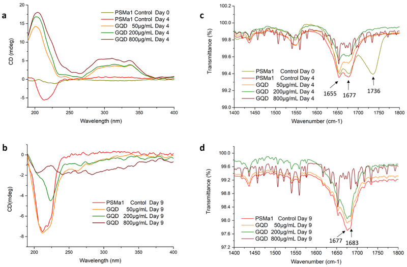
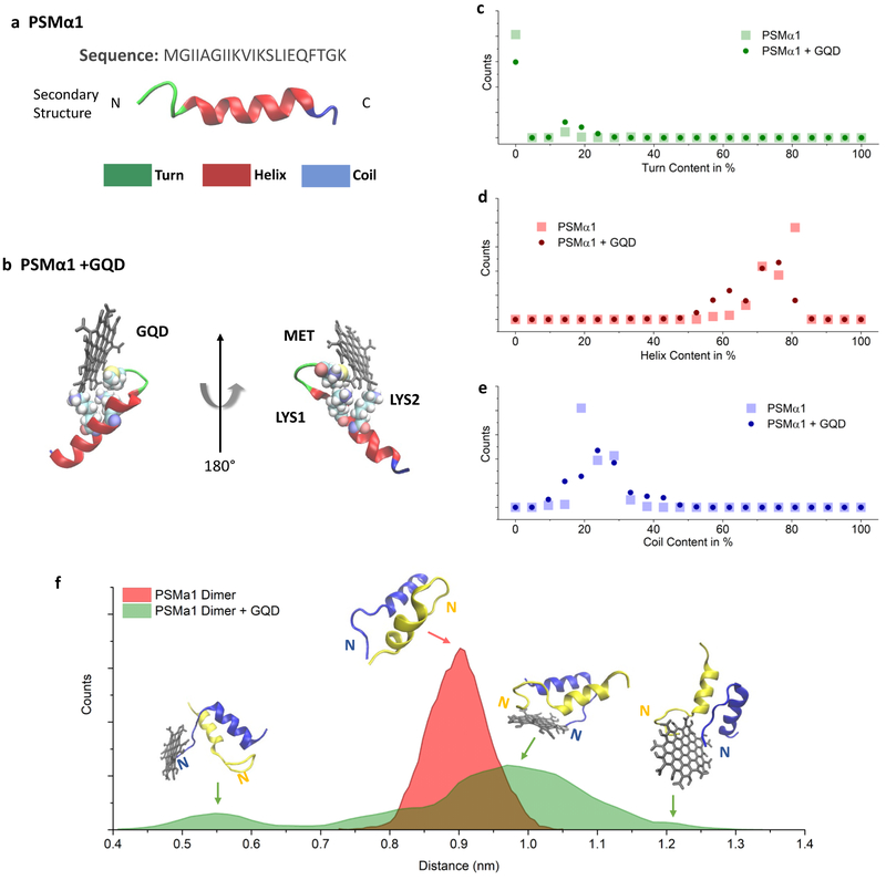
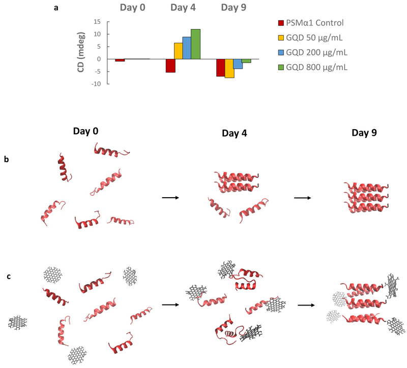
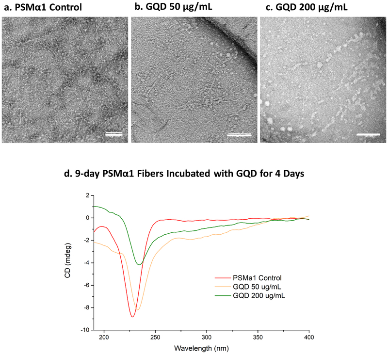
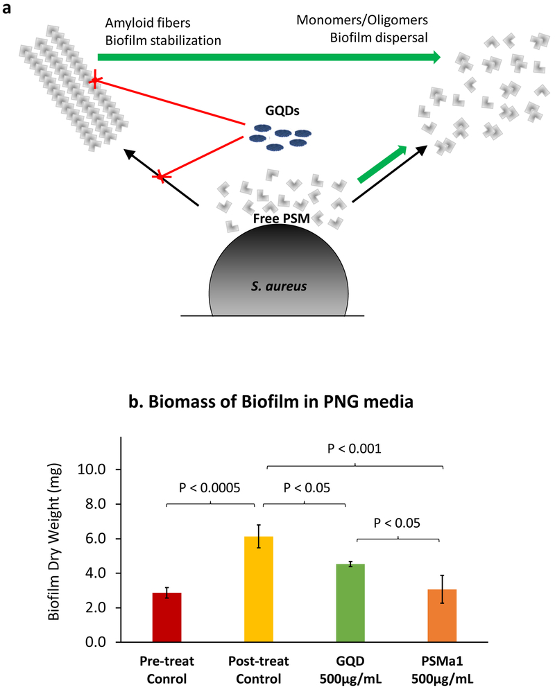
Bacterial biofilms represent an essential part of Earth's ecosystem that can cause multiple ecological, technological, and health problems. The environmental resilience and sophisticated organization of biofilms are enabled by the extracellular matrix that creates a protective network of biomolecules around the bacterial community. Current anti-biofilm agents can interfere with extracellular matrix production but, being based on small molecules, are degraded by bacteria and rapidly diffuse away from biofilms. Both factors severely reduce their efficacy, while their toxicity to higher organisms creates additional barriers to their practicality. In this paper, we report on the ability of graphene quantum dots to effectively disperse mature amyloid-rich Staphylococcus aureus biofilms, interfering with the self-assembly of amyloid fibers, a key structural component of the extracellular matrix. Mimicking peptide-binding biomolecules, graphene quantum dots form supramolecular complexes with phenol-soluble modulins, the peptide monomers of amyloid fibers. Experimental and computational results show that graphene quantum dots efficiently dock near the N-terminus of the peptide and change the secondary structure of phenol-soluble modulins, which disrupts their fibrillation and represents a strategy for mitigation of bacterial communities.

Keywords: biofilm regulation; carbon nanomaterials; extracellular matrix; functional amyloid; nanoscale biomimetics; phenol-soluble modulins; quorum sensing.


Conflict of interest statement

DECLARATION OF INTERESTS

The authors declare no competing interests.

Figures

Figure 1.
Figure 1.. Effects of GQDs on S. aureus biofilms.
(a-f) Confocal microscopy of S. aureus biofilm grown for 3 days and then treated for 1 day; stains - polysaccharide intercellular adhesin (PIA; green) and bacterial cells (blue). Biofilms grown in TSBG medium in presence of (a) 0 μg/mL (f) 50 μg/mL and (c) 500 μg/mL GQDs. Biofilms grown in PNG medium in presence of (d) 0 μg/mL (e) 50 μg/mL and (f) 500 μg/mL GQDs. Bar graphs of quantified (g) surface coverage, (h) thickness and (i) void size of biofilms analyzed from the z-stack images for each group in (a-f). (j, k) Representative SEM images and their (l) calculated surface coverage of S. aureus biofilms grown in PNG media. Scale bar: 200 μm (a-f), 50 μm (j, k).
Figure 2.
Figure 2.. TEM and CD spectra of isolated ECM from S. aureus biofilm grown in PNG and exposed to GQDs.
(a) Amyloid fibrils in composition with ECM attached to bacteria; (b) Extracted composite ECM treated with (b) 0 μg/mL and (c) 50 μg/mL GQDs for 4 days, (d) CD spectra of isolated ECM treated with 0 μg/mL, 10 μg/mL, and 50 μg/mL GQD for 4 days. Scale bars: 500 nm (a), 100 nm (b, c). Only low concentrations of GQDs were evaluated in this test due to the low concentration of ECM obtained from the isolation procedure compared to that from intact biofilms.
Figure 3
Figure 3. PSMα1 fibril formation with and without addition of GQDs.
TEM images of PSMα1 peptides after addition of 0, 50, 200, 800 μg/mL GQDs (a-d) incubated for 4 days or (e-h) 9 days. Scale bars: 100 nm.
Figure 4.
Figure 4.. Secondary structure of synthetic PSMα1 peptides and fibrils with GQDs.
(a, b) CD spectra of PSMα1 with/without addition of 50, 200, 800 μg/mL GQDs for (a) 4 days and (b) 9 days. FTIR spectra of PSMα1 with/without addition of 50, 200, 800 μg/mL GQDs for (c) 4 days and (d) 9 days. The intense signals between 1735–1740 cm−1, for control PSMα1 on Day 0, are indicative of C=O stretch of carboxylic group, which disappeared after incubation for four days in all samples. This is contributed by the minimum remains of trifluoroacetic acid (TFA) and hexafluoroisopropanol (HFIP), which cleave synthesized peptides to mono-dispersed peptide (Figure S7). This peak disappeared after 4 hours at room temperature and the remains did not change the pH of the peptide solution.
Figure 5.
Figure 5.. MD simulation of PSMα1 and GQDs.
Schemes of (a) PSMα1 and (b) PSMα1 and GQD complex, β-tiirns are shown in green, alpha helix in red and random coils in blue, (c-e) Histogram of secondary structure amounts in PSMα1 in the GQD/PSMα1 complex, (f) Histogram of the center of mass distance between two PSMα1 units showing the distance distribution with and without a GQD molecule. N: N-terminal of PSMα1; C: C-terminal of PSMα1.
Figure 6.
Figure 6.. Schematics of the effect of GQDs on PSM peptide fibrillation.
(a) The timeline of CD intensity at the typical β-sheet signal, a negative band at ≈ 218 nm indicates the process of PSM peptide fibrillation with/without addition of 50,200, 800 μg/mL GQDs. (b, c) GQDs interfere on N-terminal of PSM monomers or dimers and thereby frustrate fibrillation.
Figure 7.
Figure 7.. Effect pf GQDS on mature amyloid fibers.
PSMα1 fibers matured for 9 days were exposed to GQDs for 4 days. TEM images of PSMα1 fibers matured for 9 days (a) without GQD and with (b) 50 μg/mL and (c) 200 μg/mL GQDs were incubated for 4 more days. (d) CD spectra of PSMα1 fibers matured for 9 days with or without 50 μg/mL, and 200 μg/mL GQD for 4 more days. Scale bars: 100 nm (a-c).
Figure 8
Figure 8. Schematics of GQD mediated staphylococcal biofilm dispersal.
(a) GQDs interact with PSM peptides in two ways. First, they inhibit fibrillation of free peptides. Second the disrupt already formed fibers. These events prevent robust stabilization of the biofilm. In addition, they lead to an increase in free monomeric and oligomeric PSM peptides which trigger dispersal events, (b) Dry weights of S. aureus biofilm in PNG media treated with 500 μg/mL GQD and PSMα1.

References

    1. Hill EC; Hill GC Microbial Contamination and Associated Corrosion in Fuels, during Storage, Distribution and Use. Adv. Mater. Res. 2008, 38, 257–268.
    1. Porcelli N; Judd S Chemical Cleaning of Potable Water Membranes: A Review. Sep. Purif. Technol. 2010, 77, 137–143. - PubMed
    1. Costerton JW Bacterial Bofilms: A Common Cause of Persistent Infections. Science (80-.). 1999, 284, 1318–1322. - PubMed
    1. Castaño-Rodríguez N; Goh K-L; Fock KM; Mitchell HM; Kaakoush NO Dysbiosis of the Microbiome in Gastric Carcinogenesis. Sci. Rep. 2017, 7, 15957. - PMC - PubMed
    1. Davey ME; O'toole GA Microbial Biofilms: From Ecology to Molecular Genetics. Microbiol. Mol. Biol. Rev. 2000, 64, 847–867. - PMC - PubMed

Publication types

MeSH terms