Review
. 2019 May 13;20(5):1829-1848.
doi: 10.1021/acs.biomac.9b00228. Epub 2019 Apr 4.

Protein Assemblies: Nature-Inspired and Designed Nanostructures

Affiliations
Review

Protein Assemblies: Nature-Inspired and Designed Nanostructures

Ian W Hamley. Biomacromolecules. .

Abstract

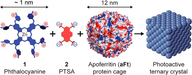
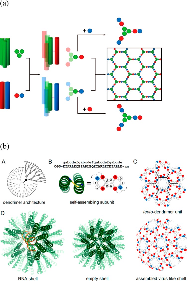
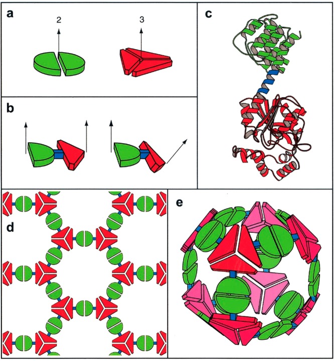
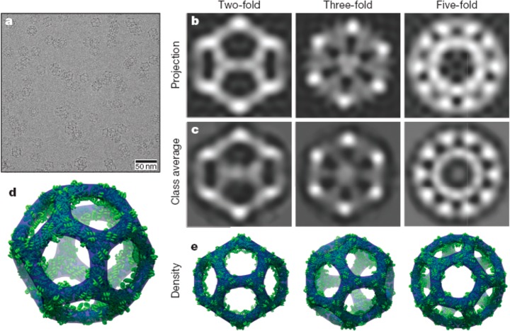
Ordered protein assemblies are attracting interest as next-generation biomaterials with a remarkable range of structural and functional properties, leading to potential applications in biocatalysis, materials templating, drug delivery and vaccine development. This Review covers ordered protein assemblies including protein nanowires/nanofibrils, nanorings, nanotubes, designed two- and three-dimensional ordered protein lattices and protein-like cages including polyhedral virus-like cage structures. The main focus is on designed ordered protein assemblies, in which the spatial organization of the proteins is controlled by tailored noncovalent interactions (including metal ion binding interactions, electrostatic interactions and ligand-receptor interactions among others) or by careful design of modified (mutant) proteins or de novo constructs. The modification of natural protein assemblies including bacterial S-layers and cage-like and rod-like viruses to impart novel function, e.g. enzymatic activity, is also considered. A diversity of structures have been created using distinct approaches, and this Review provides a summary of the state-of-the-art in the development of these systems, which have exceptional potential as advanced bionanomaterials for a diversity of applications.


Conflict of interest statement

The author declares no competing financial interest.

Figures

Figure 1
Figure 1
(a) Concept to construct Nanolego A and Nanolego B building blocks from the homotetrameric S4 protein (superoxide reductase) modified with peptides Ba and Bb (PDZ domain peptide and PDZ-binding peptide) by creating fusion proteins using the subunit C-termini (shown). (b) Pairwise linear self-assembly leads to nanofibril formation. Reproduced from ref (1) with permission of John Wiley and Sons. Published by Wiley-Blackwell. Copyright 2009 The Protein Society.
Figure 2
Figure 2
(A) Nanoring (toroid) formation using(bis-methotrexate MTX2-C9 binding to Escherichia coli dihydrofolate reductase variants with extended spacers between the two subunits (B). (C) TEM image showing nanorings assembled in a solution of 1DD-G with MTX2-C9. Reproduced with permission from ref (43). Copyright 2006 American Chemical Society.
Figure 3
Figure 3
Dimeric coiled coil peptides with a flexible (GN)x linker between the two complementary helical peptides can form extended fibrils (x = 1), triangular trimers (x = 3) or square tetramers (x = 4). Reproduced with permission from ref (14). Copyright 2012 American Chemical Society.
Figure 4
Figure 4
Nanotube structure from assembly of a designed cytochrome-based protein subunit. (a) Cryo-TEM image of a representative nanotube. (b) Helical arrangement of tetrameric proteins into a nanotube structure stabilized by zinc ion coordination at the interfaces (i-faces) shown, Zn1 for example denotes the dimer of C2-symmetric dimers forming the tetrameric building block. (c) Models for the outer (top) and inner (bottom) nanotube surfaces showing ridges and plateaus as shown side-on in panel b. Reprinted by permission from Springer Nature, ref (19). Copyright 2012 Macmillan Publishers Limited, https://www.nature.com/nchem/.
Figure 5
Figure 5
Soybean agglutinin (SBA) is a homotetrameric protein with D2 symmetry. Addition of designed ligands incorporating a galactosamine (shown) or galactopyranoside unit able to bind SBA linked to a Rhodamine B unit able to undergo π–π stacking interactions, drives protein association, leading to helical wrapping into nanotubes (scale bar = 25 nm). Reproduced with permission from ref (15). Copyright 2016 American Chemical Society.
Figure 6
Figure 6
(a) Schematic to show modification of protein RhuA with eight biotins (bR), showing two of them binding to streptavidin (S), this in turn binding to biotinylated streptavidin linkers (bbS). The C terminal (Ct) units are highlighted; these are sites for hexahistidine tagging. (b,c) Representative TEM images of aggregates of bR and bR.S4 on lipid monolayers. The scale bars indicate 200 nm. From ref (73). Reprinted with permission from AAAS. http://science.sciencemag.org/content/302/5642/106.
Figure 7
Figure 7
Modification of protein interfaces (mutations marked by purple spheres) to favor small oligomers. (a) Crystal structure of C2-symmetric UroA tetramer showing the 2-fold molecular symmetry axis (red) and four local 2-fold axes relating the cores (black lines). (b) Octamer formed by dimerization of RhuA dimer with D4 symmetry. (c) RhuB octamer with C2 symmetry. (d) Negative stain TEM image of RhuE showing assembly of fibers, the inset scale bar shows a RhuA octamer to scale. (e) Native mycobacterial porin, with the deleted membrane-immersion part indicated by the box, giving MypA. (f) D8-symmetric assembly of two MypA molecules (top and bottom rings). The positions of the 52-residue deletions are marked by red spheres. From ref (74). Reprinted with permission from AAAS. http://science.sciencemag.org/content/319/5860/206.
Figure 8
Figure 8
Distinct 2D lattices formed by the indicated RhuA mutants. (i) Low magnification TEM images, (ii) High magnification TEM images. (iii) Fourier transforms of images in column ii. (iv) Reconstructed 2D images from the Fourier transforms. (v) Structural models based on iv. Reprinted by permission from Springer Nature, ref (20). Copyright 2016 Macmillan Publishers Limited, https://www.nature.com/.
Figure 9
Figure 9
Galactose-based ligand-driven assembly of LecA homotetrameric proteins. (a) Structure of the LecA tetramer (protein data bank pdb id 4LKD). (b) Chemical structures of ligands RnG (n = 1 to 5) and R4M. (c) Illustration of dimerization. (d) Possible arrangements of LecA/RnG giving rise to different 1D and 2D nanostructures. Reproduced from ref (16) with permission of John Wiley and Sons. Copyright 2017 Wiley-VCH Verlag GmbH & Co. KGaA, Weinheim.
Figure 10
Figure 10
Fusion of protein assembly elements leads to 1D and 2D superstructures (termed crysalins). Left column: Schematic of assembly units showing symmetry elements. Second column: Protein/peptide components incorporated in fusion proteins. Third column: Designed structures. Right column: TEM images of observed structures. Reprinted by permission from Springer Nature, ref (4). Copyright 2011 Macmillan Publishers Limited, https://www.nature.com/nnano/.
Figure 11
Figure 11
Creation of designed 2D lattices by protein design. (A, F, K) targeted lattices with (inset) protein subunit arrangements, (B, G, L) models for designed proteins packed into the lattices shown above, (C, H, M) designed interface structure for the corresponding lattice structure in the same column, (D, I, N) cryo-TEM images of expressed protein lattices (white scale bars = 50 nm, black scale bars = 5 nm), with inset Fourier transforms. (E, J, O) Calculated projection maps (14 or 15 Å resolution) with overlaid protein designs shown on the right of each image. From ref (75). Reprinted with permission from AAAS. http://science.sciencemag.org/content/348/6241/1365.
Figure 12
Figure 12
2D lattices with p6 symmetry from a modified hexameric S. typhimurium STM4215 protein TTM. (a) Ribbon structure showing native hexameric structure. (b) A dimer linked by an introduced short six-residue sequence (blue) with Rosetta designed modified interfaces (shown in black). (c) Top and side views of the expected 2D lattice with individual TTM dimers shown in different colors. (d) TEM image after incubating a protein sample with CaCl2 (inset: Fourier transform image showing hexagonal symmetry). Reproduced with permission from ref (76). Copyright 2015 American Chemical Society.
Figure 13
Figure 13
Design of a honeycomb lattice from homotrimeric coiled coils. (a) Schematic of the p6 structure along with lattice symmetry elements, the parameters θ and R adjusted in the peptide design are shown in the bottom scheme. (b) Two layers of the peptides showing H-bonding at the interlayer interface facilitated by fixing the unit cell length c. (c) Electron density map from a single crystal structure (bottom) compared to a model structure (top). Reproduced from ref (24) with permission of the authors.
Figure 14
Figure 14
(a) Schematic showing a ferritin C4-symmetry axis with pore, based on assembly of subunits shown on left with E-helix highlighted, (b) Surface charges on ferritin–orange spheres indicates negative charges and black spheres are positive charges, (c) Showing strategy to expand pore size by E-helix deletion from H-1 subunits in reconstructed mature soybean seed ferritin (rmSSF). The expanded pore size enables ingress of poly(l-lysine) which links proteins into a square array via electrostatic interactions. From Chemical communications by Royal Society of Chemistry (Great Britain). Republished with permission of Royal Society of Chemistry, from ref (25). Copyright 2014; permission conveyed through Copyright Clearance Center, Inc.
Figure 15
Figure 15
2D and 3D assemblies by aromatic substitution within the 24-subunit ferritin protein cage structure. (a) Schematic of ferritin structure showing C4 rotation axes and substitution sites near these axes, leading to 2D and 3D lattices depending on the aromatic residue substituted at Glu162. (b) Showing one of the 4-fold symmetry axes of human H-chain ferritin (pdb file 2FHA). (c) Reconstructed image from FFT of a TEM image of a 2D oblique (rhombic) lattice for the phenylalanine substituted protein assembly. (d) SAXS pattern and one-dimensional intensity profile with indexed reflections corresponding to a simple cubic packed 3D structure (shown) for the tyrosine-substituted protein assembly. Parts a, c, d reproduced with permission from ref (18). Copyright 2018 American Chemical Society.
Figure 16
Figure 16
Concept of swellable protein-embedded polymer hydrogel crystals. (a–c) Showing fcc packing in ferritin crystals (Protein Data bank Identifier, pdb id 6B8F). (d) Ca2+-mediated interactions leading to the packing of ferritin proteins in the crystal lattice. (e) Schematic of polymerization around the ferritin lattice scaffold to produce a reversibly swellable hybrid polymer–protein crystal hydrogel structure. Reprinted by permission from Springer Nature, ref (80). Copyright 2018 Macmillan Publishers Limited, https://www.nature.com/.
Figure 17
Figure 17
Complexes form between a zinc phthalocyanine derivative 1 and the tetra-anionic pyrene derivative PTSA (1,3,6–8-pyrene tetra-sulfonic acid) 2. These complexes bind to anionic patches on the apoferritin protein surface, leading to the formation of cubic crystals which retain the photoactivity of the phthalocyanine dye. Reproduced with permission from ref (81). Copyright 2015 American Chemical Society.
Figure 18
Figure 18
(a) Schematic for coiled coil peptide assembly designed to self-assemble into a honeycomb lattice (which is observed to curve into a cage structure). Left: a homotrimeric coiled coil is linked via cysteine disulfide cross-linking to a homodimeric coiled coil. Mixing of either the top building block (center, green and red) termed Hub A with coiled coil module B (basic coil peptide, blue) or Hub B (center bottom 3-arm structure, green and blue) and module A (acidic coil peptide, red) leads to the formation of a honeycomb lattice (right). (b) Design of a dendrimer-like coiled coil peptide which forms a cage structure. (A) Dendrimer architecture, (B) cysteine-linked (yellow connector) coiled coil dimer; red and blue circles indicate glutamate and arginine residues, respectively. (c) Expected honeycomb lattice, (D) model for RNA-filled capsule, empty shell and observed virus-like cage structure. Part a from ref (87). Reprinted with permission from AAAS. http://science.sciencemag.org/content/340/6132/595. Part b reproduced with permission from ref (26). Copyright 2016 American Chemical Society.
Figure 19
Figure 19
Fusion protein design. (a) Proteins with different subunit symmetries (here 2-fold and 3-fold rotation symmetry). (b) Fusion of two proteins (showing two possible geometries). (c) A ribbon diagram showing an example of a fusion construct where red and green proteins are linked by a short α-helix (blue). The fusion requires one protein to have an initial α-helix domain, the other protein must have a terminal α-helix. (d) Schematic of a 2D honeycomb lattice that assembles from flat fusion dimers. (e) Schematic of a cage structure formed when the two proteins are twisted, as shown in part b, right. Reproduced from ref (6). Copyright 2001 National Academy of Sciences, U.S.A.
Figure 20
Figure 20
Icosahedral protein cages. (a) Low-magnification cryo-TEM image showing cages in different projections. (b) Back-projections of structure along different symmetry axes based on the model. (c) Class averages from cryo-TEM images (bottom). (d) Three-dimensional model of the icosahedral structure. (e) Projections corresponding to images in panels b and c. Reprinted by permission from Springer Nature, ref (8). Copyright 2016 Macmillan Publishers Limited, https://www.nature.com/.
Figure 21
Figure 21
(a) Design of a fusion protein with appropriate oriented symmetry axes based on a trimeric protein (green) linked to a dimeric domain (orange) via a four-residue helical linker (blue). (b) Intended 24-subunit cubic cage structure with octahedral symmetry, the 3-fold symmetry axes (cyan) and 2-fold symmetry axes (magenta) of a cube being shown on the right. Reprinted by permission from Springer Nature, ref (10). Copyright 2014 Macmillan Publishers Limited, https://www.nature.com/nchem/.
Figure 22
Figure 22
Polyhedral peptide nanoparticles based on (a) building block comprising two linked coiled-coil peptides designed to form pentamers (green) or trimers (blue), with (b) models for their assembly into icosahedral particles. Top: Nanoparticle containing 60 peptides. Bottom: nanoparticle containing 180 peptides. Reprinted from refs (97, 98). Copyright 2006 and 2011, with permission from Elsevier.
Figure 23
Figure 23
(a, b) Fusion protein from a designed four-helix dimer and a trimer from T4 phage fibritin. (c) Possible assemblies expected for the fusion protein, which are based on multiples of 6-mers. Reproduced with permission from ref (11). Copyright 2015 American Chemical Society.
Figure 24
Figure 24
Incorporation of alcohol dehydrogenase into the pET expression vector for bacteriophage P22 produces the AlhD-SP conjugate (red, with C-terminal truncated scaffold protein shown in yellow), and coassembly with the coat protein shown in blue leads to assembly of virus-like particles shown on the right, decorated with enzymes on the interior with model enzymatic activity shown (NAD: nicotinamide adenine dinucleotide). Reproduced with permission from ref (12). Copyright 2012 American Chemical Society.

References

    1. Usui K.; Maki T.; Ito F.; Suenaga A.; Kidoaki S.; Itoh M.; Taiji M.; Matsuda T.; Hayashizaki Y.; Suzuki H. Nanoscale elongating control of the self-assembled protein filament with the cysteine-introduced building blocks. Protein Sci. 2009, 18 (5), 960–969. 10.1002/pro.106. - DOI - PMC - PubMed
    1. Chou T.-F.; So C.; White B. R.; Carlson J. C. T.; Sarikaya M.; Wagner C. R. Enzyme nanorings. ACS Nano 2008, 2, 2519–2525. 10.1021/nn800577h. - DOI - PMC - PubMed
    1. Moll D.; Huber C.; Schlegel B.; Pum D.; Sleytr U. B.; Sara M. S-layer-streptavidin fusion proteins as template for nanopatterned molecular arrays. Proc. Natl. Acad. Sci. U. S. A. 2002, 99 (23), 14646–14651. 10.1073/pnas.232299399. - DOI - PMC - PubMed
    1. Sinclair J. C.; Davies K. M.; Venien-Bryan C.; Noble M. E. M. Generation of protein lattices by fusing proteins with matching rotational symmetry. Nat. Nanotechnol. 2011, 6 (9), 558–562. 10.1038/nnano.2011.122. - DOI - PubMed
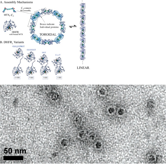
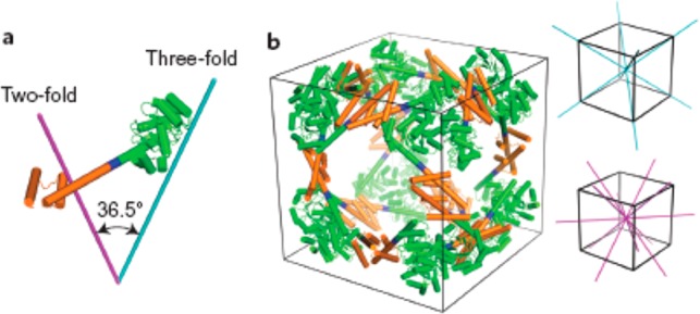
    1. Sciore A.; Su M.; Koldewey P.; Eschweiler J. D.; Diffley K. A.; Linhares B. M.; Ruotolo B. T.; Bardwell J. C. A.; Skiniotis G.; Marsh E. N. G. Flexible, symmetry-directed approach to assembling protein cages. Proc. Natl. Acad. Sci. U. S. A. 2016, 113 (31), 8681–8686. 10.1073/pnas.1606013113. - DOI - PMC - PubMed

MeSH terms

Substances