Clinical Trial
. 2019 Mar 26;14(3):e0214202.
doi: 10.1371/journal.pone.0214202. eCollection 2019.

Autoantibodies are present before the clinical diagnosis of systemic sclerosis

Affiliations
Clinical Trial

Autoantibodies are present before the clinical diagnosis of systemic sclerosis

Peter D Burbelo et al. PLoS One. .

Abstract

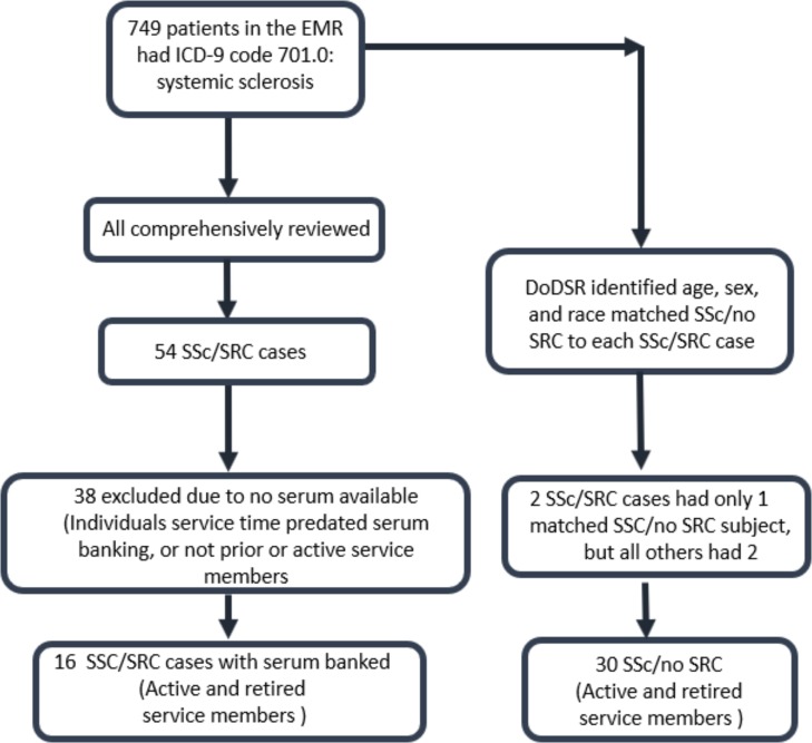
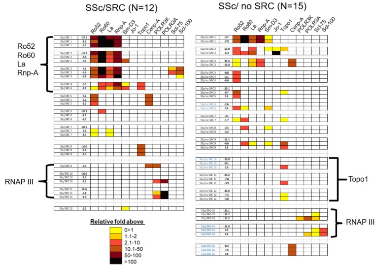
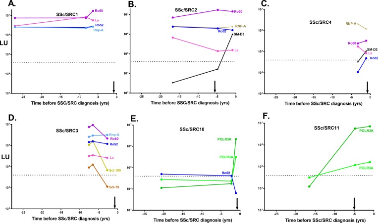
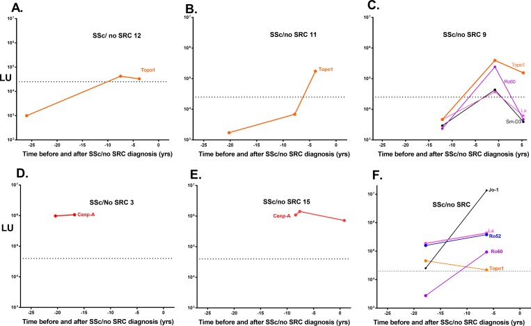
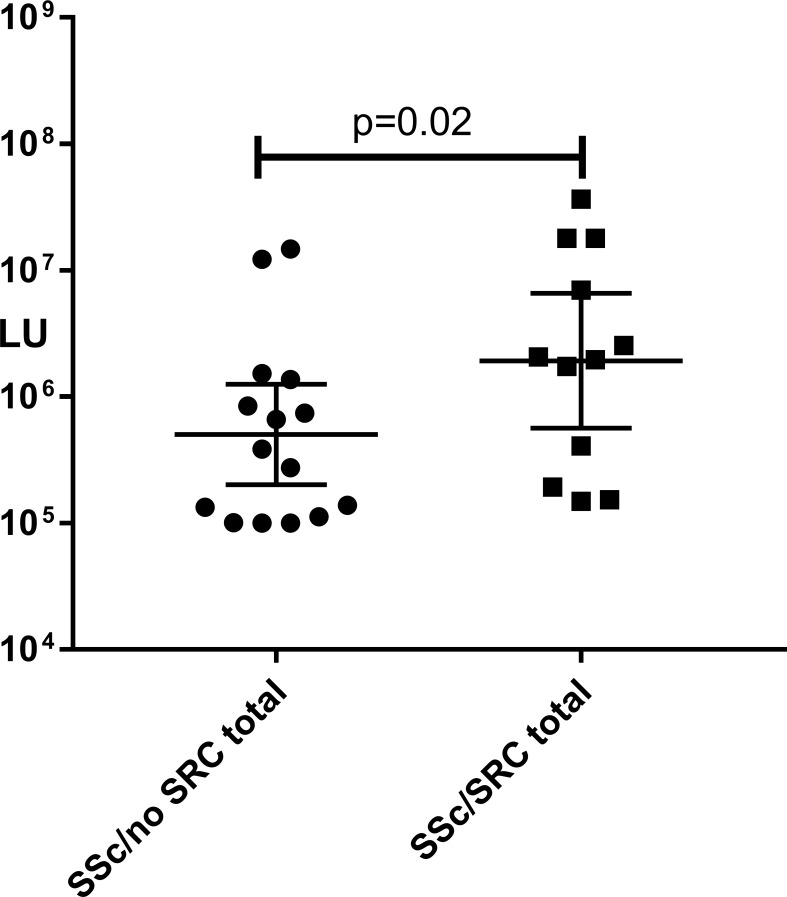
Systemic sclerosis (SSc) is a heterogeneous autoimmune disorder associated with vascular dysfunction and fibrotic changes in the skin, vasculature and internal organs. Although serologic abnormalities are an important diagnostic tool for SSc, little is known about whether autoantibodies precede clinical diagnosis. Here we investigated the presence of autoantibodies before SSc diagnosis and assessed whether certain autoantibodies might associate with the future onset of scleroderma renal crisis (SRC), a potentially fatal complication of the disease. Using the Department of Defense Serum Repository, autoantibodies were analyzed from archived, prospectively collected, longitudinal serum samples from sixteen individuals with SRC (SSc/SRC) and thirty cases of SSc without SRC (SSc/no SRC), matched for age, sex, and race. Seventy five percent (12/16) of the SSc/SRC and 40% (12/30) of the SSc/no SRC were seropositive for at least one autoantibody prior to clinical diagnosis (up to 27.1 years earlier, mean = -7.4 years). Although both disease groups demonstrated a heterogeneous immunoreactivity profile against the autoantigen panel, the SSc/SRC subjects showed two enriched clusters with one featuring elevated levels of autoantibodies against Ro52 and/or Ro60 and another with high levels of immunoreactivity against the RNA polymerase complex. Consistent with larger spectrum of immunoreactivity and the elevated levels of autoantibodies in SSc/SRC, the total response against the autoantigen panel from the last time point of the seropositive subjects revealed that the SSc/SRC cohort harbored higher antibody levels (p = 0.02) compared to SSc/no SRC. Overall, our findings demonstrate that relevant seropositive autoantibodies often precede the clinical diagnosis of SSc/no SRC and SSc/SRC.


Conflict of interest statement

The authors have declared that no competing interests exist.

Figures

Fig 1
Fig 1. Flow-chart for selection of the SSc/SRC and SSc/no SRC cases.
As described in the material and methods, screening of the military electronic medical records between 2005–2016 for Systemic Sclerosis was initially preformed. Following comprehensive review, 54 cases of SS/SRC were identified, of which only 16 had available serum samples. Thirty additional SSc/no SRC cases were then identified that were matched for age, gender and race with the SS/SRC group.
Fig 2
Fig 2. Serum autoantibody levels in the SSc/SRC and SSc/no SRC cohort.
Autoantibody measurements were made by LIPS against the 12 autoantigens in the 121 blinded serum samples corresponding to a total of 16 SRC and 30 SSc/no SRC cases. The Y axis reflects the antibody levels in LU determined by LIPS. The blue line is the cut-off value for each antigen. As shown by the numbers in blue, the most prevalent autoantibodies were against Ro52, while the least common were against the Jo-1 autoantigen.
Fig 3
Fig 3. Heatmap analysis of autoantibodies in the SSc/SRC and SSc/no SRC cases.
Heatmap analysis of autoantibody responses are shown in the 12 seropositive SSc/SRC and 15 seropositive SSc/no SRC subjects. For each case, the time in years before (-) or after (+) initial systemic sclerosis diagnosis is denoted in the column on the left. Each group of rows represents the autoantibody profile in a single case, in which the blue colored codes represent SSc cases with autoantibodies detected after diagnosis. Color coding denotes relative antibody levels above the baseline cut-off value and the clear boxes represent seronegative responses with the autoantigens in a given subject. As shown by the key, seropositive autoantibody levels in the subjects ranged from low levels (yellow) to extremely high autoantibody levels (black). Based on the patterns that emerged, the SSc/no SRC and SSc/SRC subjects were then manually segregated into three autoantibody clusters for Ro6o/Ro52/La/Rnp-A, Topo1 and RNAP III.
Fig 4
Fig 4. Representative autoantibody profiles seen in SSc/SRC cases before disease diagnosis.
Representative plots illustrating autoantibody levels in six SSc/SRC subjects before disease diagnosis. The X-axis denotes the time in years before diagnosis of SSc/SRC (time 0). The approximate time of SSc/no SRC diagnosis preceded SSc/SRC and is denoted by the black vertical arrow. The left Y axis represents the autoantibody levels in LU and the dotted line represent the approximate cut-off value for the autoantigens.
Fig 5
Fig 5. Representative autoantibody profiles seen before diagnosis of SSc/no SRC.
Representative plots illustrating autoantibody levels in the six SSc/ no SRC subjects before diagnosis of the disease. The X-axis denotes the time in years before diagnosis. The left Y axis represents the scale of the autoantibody levels in LU and the dotted line represent the cut-off values for the antigens. All seropositive antibody responses against the autoantigen panel are shown.
Fig 6
Fig 6. Higher autoantibody levels in SSc/SCR compared to SSc/no SRC cases.
The scatter plot graphs represent the total antibody response in individual seropositive SSc/SRC and SSc/no SRC cases. This analysis was accomplished by summing up the autoantibody values against the twelve autoantigens from the last time point of the seropositive subjects. The P values were calculated using the Mann-Whitney U test.

References

    1. Gabrielli A, Avvedimento EV, Krieg T. Scleroderma. N Engl J Med. 2009;360(19):1989–2003. 10.1056/NEJMra0806188 . - DOI - PubMed
    1. Denton CP, Khanna D. Systemic sclerosis. Lancet. 2017;390(10103):1685–99. Epub 2017/04/18. 10.1016/S0140-6736(17)30933-9 . - DOI - PubMed
    1. LeRoy EC, Black C, Fleischmajer R, Jablonska S, Krieg T, Medsger TA Jr., et al. Scleroderma (systemic sclerosis): classification, subsets and pathogenesis. J Rheumatol. 1988;15(2):202–5. Epub 1988/02/01. . - PubMed
    1. Pakozdi A, Nihtyanova S, Moinzadeh P, Ong VH, Black CM, Denton CP. Clinical and serological hallmarks of systemic sclerosis overlap syndromes. J Rheumatol. 2011;38(11):2406–9. Epub 2011/08/17. 10.3899/jrheum.101248 . - DOI - PubMed
    1. Mouthon L, Bussone G, Berezne A, Noel LH, Guillevin L. Scleroderma renal crisis. J Rheumatol. 2014;41(6):1040–8. 10.3899/jrheum.131210 . - DOI - PubMed

Publication types