. 2019 May;49(5):320-329.
doi: 10.2519/jospt.2019.8793. Epub 2019 Mar 26.

Are Magnetic Resonance Imaging Technologies Crucial to Our Understanding of Spinal Conditions?

Are Magnetic Resonance Imaging Technologies Crucial to Our Understanding of Spinal Conditions?

Rebecca J Crawford et al. J Orthop Sports Phys Ther. 2019 May.

Abstract

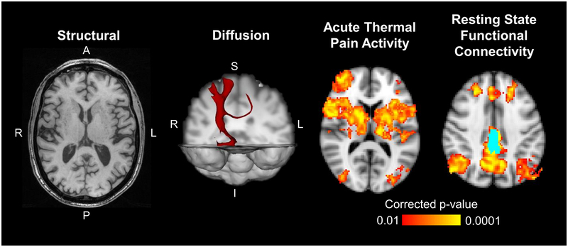
Persistent spinal (traumatic and nontraumatic) pain is common and contributes to high societal and personal costs globally. There is an acknowledged urgency for new and interdisciplinary approaches to the condition, and soft tissues, including skeletal muscles, the spinal cord, and the brain, are rightly receiving increased attention as important biological contributors. In reaction to the recent suspicion and questioned value of imaging-based findings, this paper serves to recognize the promise that the technological evolution of imaging techniques, and particularly magnetic resonance imaging, is allowing in characterizing previously less visible morphology. We emphasize the value of quantification and data analysis of several contributors in the biopsychosocial model for understanding spinal pain. Further, we highlight emerging evidence regarding the pathobiology of changes to muscle composition (eg, atrophy, fatty infiltration), as well as advancements in neuroimaging and musculoskeletal imaging techniques (eg, fat-water imaging, functional magnetic resonance imaging, diffusion imaging, magnetization transfer imaging) for these important soft tissues. These noninvasive and objective data sources may complement known prognostic factors of poor recovery, patient self-report, diagnostic tests, and the "-omics" fields. When combined, advanced "big-data" analyses may assist in identifying associations previously not considered. Our clinical commentary is supported by empirical findings that may orient future efforts toward collaborative conversation, hypothesis generation, interdisciplinary research, and translation across a number of health fields. Our emphasis is that magnetic resonance imaging technologies and research are crucial to the advancement of our understanding of the complexities of spinal conditions. J Orthop Sports Phys Ther 2019;49(5):320-329. Epub 26 Mar 2019. doi:10.2519/jospt.2019.8793.

Keywords: MRI; muscle; pain; soft tissues; spinal cord; spine.


Conflict of interest statement

CONFLICT OF INTEREST STATEMENT

RC reports no conflict of interest

MF reports no conflict of interest

KW reports funding from the National Institutes of Health (NIH)

AC reports funding from NIH

JE reports funding from NIH; Advisory member for the Board of Directors, JOSPT; Editorial board member for JOSPT, Musculoskeletal Science & Practice; Advisory Board member the journal, Spine. Honoraria, conference travel, and accommodation for speaking engagements.

Figures

Figure 1a-b -
Figure 1a-b -
Example of axial T2-weigthed lumbar paraspinal muscle morphology and composition measurements. a) illustrates the cross-sectional area measurements of the psoas (1,2), quadratus lumborum (3,4), multifidus (5,6) and erector spinae (7, 8), and also demonstrates current methodological differences in ROI definitions for the erector spinae muscle; the fat-filled “tent-region” under the lumbosacral fascia (posteriorly) was included in (7) and excluded in (8). b) illustrates the functional cross-sectional area (in green) representing the area of lean muscle mass (excluding fatty infiltration) of the multifidus muscle using a thresholding technique.
Figure 2 -
Figure 2 -
Example of axial fat-only images examining composition of the multifidus and semispinalis cervicis traversing the cervical spine. a) illustrates the cross-sectional area measure of the multifidus and semispinalis muscles in the mid-cervical region (C3) and b) illustrates the cross-sectional area measure of the multifidus and semispinalis muscles in the lower-cervical region (C7). c) illustrates the propagated volume of the multifidus and semispinalis cervicis spanning those vertebral levels.
Figure 3 –
Figure 3 –
Example of Magnetization Transfer Contrast
Figure 4-
Figure 4-
Example structural, diffusion, magnetization transfer, and functional axial cervical spinal cord images are shown. The structural image was acquired using a multi-echo gradient-echo sequence, which provides high white matter to gray matter contrast. In the diffusion image, the principle direction of diffusion in the spinal cord is through the axial plane as indicated by the colored lines. Magnetization transfer imaging can be used to calculate the magnetization transfer ratio (MTR), which provides a measure of tissue macromolecule content. The functional images show group average activation from an acute thermal pain stimulus applied to the right ventral forearm and group average connectivity to the C7 right anterior horn (light blue). Green = spinal cord gray matter.
Figure 5-
Figure 5-
Example structural, diffusion, and functional brain images are shown. The axial structural image was acquired using 3D MPRAGE T1-weighted gradient-echo sequence and can provide morphometric properties of the gray matter. The diffusion example shows a 3D tractography map using the right ventral posterolateral nucleus of the thalamus as a seed. The axial functional images show average group activation from an acute thermal pain stimulus applied to the lower back and group average connectivity to the bilateral posterior cingulate cortices (light blue).

References

    1. Abbott R, Peolsson A, West J, Elliott JM, Aslund U, Karlsson A, Leinhard O. The qualitative grading of muscle fat infiltration in whiplash using fat and water magnetic resonance imaging. Spine J 2018;18(5):717–725. - PMC - PubMed
    1. Alschuler KN, Kratz AL, Ehde DM. Resilience and vulnerability in individuals with chronic pain and physical disability. Rehabil Psychol 2016;61(1):7–18. - PMC - PubMed
    1. Anderson SE, Boesch C, Zimmermann H, Busato A, Hodler J, Bingisser R, Ulbrich EJ, Nidecker A, Buitrago-Tellez CH, Bonel HM, Heini P, Schaeren S, Sturzenegger M. Are There Cervical Spine Findings at MR Imaging That Are Specific to Acute Symptomatic Whiplash Injury? A Prospective Controlled Study with Four Experienced Blinded Readers. Radiology 2012;262(2):567–575. - PubMed
    1. Apkarian AV, Bushnell MC, Treede RD, Zubieta JK. Human brain mechanisms of pain perception and regulation in health and disease. Eur J Pain 2005;9(4):463–484. - PubMed
    1. Apkarian AV, Sosa Y, Sonty S, Levy RM, Harden RN, Parrish TB, Gitelman DR. Chronic back pain is associated with decreased prefrontal and thalamic gray matter density. J Neurosci 2004;24(46):10410–10415. - PMC - PubMed

MeSH terms