Review
. 2019 Mar 29;9(3):180249.
doi: 10.1098/rsob.180249.

Protein damage, ageing and age-related diseases

Affiliations
Review

Protein damage, ageing and age-related diseases

Anita Krisko et al. Open Biol. .

Abstract

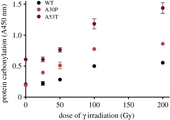
Ageing is considered as a snowballing phenotype of the accumulation of damaged dysfunctional or toxic proteins and silent mutations (polymorphisms) that sensitize relevant proteins to oxidative damage as inborn predispositions to age-related diseases. Ageing is not a disease, but it causes (or shares common cause with) age-related diseases as suggested by similar slopes of age-related increase in the incidence of diseases and death. Studies of robust and more standard species revealed that dysfunctional oxidatively damaged proteins are the root cause of radiation-induced morbidity and mortality. Oxidized proteins accumulate with age and cause reversible ageing-like phenotypes with some irreversible consequences (e.g. mutations). Here, we observe in yeast that aggregation rate of damaged proteins follows the Gompertz law of mortality and review arguments for a causal relationship between oxidative protein damage, ageing and disease. Aerobes evolved proteomes remarkably resistant to oxidative damage, but imperfectly folded proteins become sensitive to oxidation. We show that α-synuclein mutations that predispose to early-onset Parkinson's disease bestow an increased intrinsic sensitivity of α-synuclein to in vitro oxidation. Considering how initially silent protein polymorphism becomes phenotypic while causing age-related diseases and how protein damage leads to genome alterations inspires a vision of predictive diagnostic, prognostic, prevention and treatment of degenerative diseases.

Keywords: age-related diseases; ageing; protein damage.


Conflict of interest statement

We declare we have no competing interests.

Figures

Figure 1.
Figure 1.
Emergence of phenotypes of oxidatively damaged proteins in relation to cellular proteostasis (from [36]). Canonical pathways of protein synthesis and folding are represented in blue. The sources and causes of oxidative protein damage are in red. Total oxidative proteome damage is a function of both ROS level and the fraction of misfolded proteins representing effective target size for protein oxidation. In addition to rare mutations resulting from malfunction of damaged proteins, cellular phenotype is determined by the extent of oxidative proteome damage and the susceptibility of individual proteins to such damage.
Figure 2.
Figure 2.
Fidelity of translation determines the target size of proteome oxidation. Protein carbonylation (measured by ELISA-based assay) was induced by increasing doses of UVC radiation up to the saturation level. Saturation level represents the proteome target size for oxidative damage and correlates with spontaneous protein oxidation of non-irradiated cells. Streptomycin (not shown) and the rpsD (rpsD14) mutant were used to increase translational error rates. rpsL is rpsL141 mutant characterized by a reduced translational error rate. Escherichia coli was harvested from LB medium in mid-exponential growth stage, resuspended in 10 mM PBS, pH 7.4, and irradiated on ice by increasing doses of UV. Cells were collected and washed prior to protein extraction in 10 mM PBS, pH 7.4, in the presence of 1 mg ml−1 of lysozyme and protease inhibitors (Roche). Protein concentration was determined using the Bradford assay and the total protein carbonylation was measured as described previously [9].
Figure 3.
Figure 3.
Competitive antagonism between protein folding and oxidation. Selection for functional longevity of proteins led to the evolution of oxidation-resistant structures. Imperfections in native structure arise from random biosynthetic and folding errors and from silent mutations (polymorphisms). Native folding prevents oxidation and oxidation precludes correct folding. Oxidized proteins either malfunction or lose function by aggregation or proteolysis. In particular cases, oxidation could change protein function (i.e. ‘malfunction’ can become a gain of function, including the ‘non-self’ antigenicity). In all cases, there are functional (phenotypic) consequences of protein damage via direct or cascading (snowballing) effects that are reversible by antioxidants (i.e. phenotypes of misfolding are due to oxidative damage more than to misfolding itself [9,10]).
Figure 4.
Figure 4.
Protein carbonylation correlates with death due to radiation and living. (I) Radiation: The plots on the figure are schematized versions of published data [30,31]. Curves marked by A represent ‘standard’ species (E. coli, C. elegans); B represents evolved radiation-resistant E. coli and bdelloid rotifer Adineta vaga and C extremely radiation-resistant species, D. radiodurans. (II) Age: Protein carbonylation and survival versus age. A, B and C can be nematode, mouse and human cells. The crossings between A, B and C are schematized and are not in proportion on x-axis (from [36]).
Figure 5.
Figure 5.
Protein aggregate appearance in budding yeast follows the Gompertz law of mortality. (a) The replicative lifespan is presented as the number of buds produced by individual mother cells. The number of cells is 103 and 122 for the wild-type and petite (ρ0), respectively. The data shown are pooled from two independent experiments for each strain. Significance of the results was tested with log-rank test, p < 0.05. Replicative lifespan was measured as described previously [43]. (b) The figure displays the increase in the fraction of cells bearing at least one aggregate. (λ) and (μ) parameters from the Gompertz equation are indicated on the figure. (c) For illustration, spinning disc confocal microscopy images displaying representative images from different stages of lifespan (on the microscope slide, 30°C on YPD-rich medium). The exact time (min) at which a snapshot was taken is indicated on each picture. The white line represents 8 µm. See §5 for the description of methods related to this figure.
Figure 6.
Figure 6.
A small fraction of human liver proteome is sensitive to protein carbonylation. (a) A 2D-Oxi-DIGE ‘carbonylome’ of cell extracts from a human liver biopsy displays protein spots (in green) and carbonyls (in red). (b) A biopsy of a hepatocarcinoma within the same liver dispays increased cellular protein carbonylation as was found with 58 human tumours without exception. The row of seven spots in the middle of the left gel corresponds to the isoforms of the same protein with increasing negative charges due to increasing phosphorylation and carbonylation, underpinning the likely interference of different PTM. (Provided by Fernando A. Martin and Romain Ladouce, MedILS proteomic platform.)
Figure 7.
Figure 7.
Wild-type version of α-synuclein is characterized by the highest oxidation resistance. The plot displays the differential resistance to γ radiation of human α-synuclein protein isomorphs. All three proteomorphs of α-synuclein were purchased from rPeptide. Purified proteins in PBS were exposed to increasing doses of γ radiation on ice and their carbonylation level was measured as previously described [9].
Figure 8.
Figure 8.
Susceptibility of proteoforms to oxidative modifications determines the onset of their oxidation over time and leads to their functional decline. Red dots represent oxidation events. Variants of the same protein (proteoforms) characterized by stable tertiary structure are intrinsically more resistant to oxidative damage and will experience oxidation with a longer delay, compared with disordered/more flexible proteoforms. m1, m2 and m3 stand for three proteoforms (as an interpretation of data in figure 6) and their intrinsic oxidability is symbolized by the drawings above the figure.

References

    1. Kirkwood TB. 2005. Understanding the odd science of aging. Cell 120, 437–447. (10.1016/j.cell.2005.01.027) - DOI - PubMed
    1. Kirkwood TB, Kowald A. 2012. The free-radical theory of ageing—older, wiser and still alive: modelling positional effects of the primary targets of ROS reveals new support. Bioassays 34, 692–700. (10.1002/bies.201200014) - DOI - PubMed
    1. Watson J. 2013. Oxidants, antioxidants and the current incurability of metastatic cancers. Open Biol. 3, 120144 (10.1098/rsob.120144) - DOI - PMC - PubMed
    1. Khelaifia S, Lagier JC, Nkamga VD, Guilhot E, Drancourt M, Raoult D. 2016. Aerobic culture of methanogenic archaea without an external source of hydrogen. Eur. J. Clin. Microbiol. Infect. Dis. 35, 985–991. (10.1007/s10096-016-2627-7) - DOI - PubMed
    1. Tan HY, Wang N, Li S, Hong M, Wang X, Feng Y. 2016. The reactive oxygen species in macrophage polarization: reflecting its dual role in progression and treatment of human diseases. Oxid. Med. Cell Longev. 2016, 2795090 (10.1155/2016/2795090) - DOI - PMC - PubMed

Publication types

LinkOut - more resources