. 2019 Jul 15:93:97-110.
doi: 10.1016/j.actbio.2019.03.046. Epub 2019 Mar 23.

An in vitro and in vivo comparison of cartilage growth in chondrocyte-laden matrix metalloproteinase-sensitive poly(ethylene glycol) hydrogels with localized transforming growth factor β3

Affiliations

An in vitro and in vivo comparison of cartilage growth in chondrocyte-laden matrix metalloproteinase-sensitive poly(ethylene glycol) hydrogels with localized transforming growth factor β3

Margaret C Schneider et al. Acta Biomater. .

Abstract

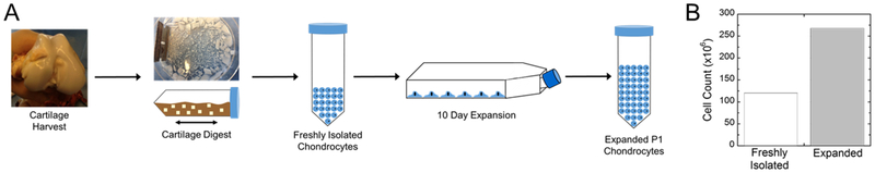
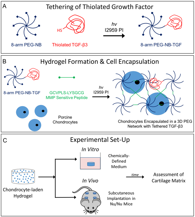
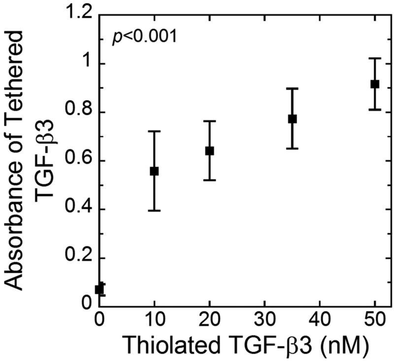
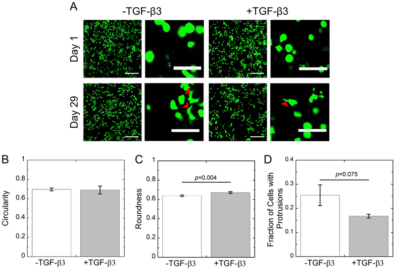
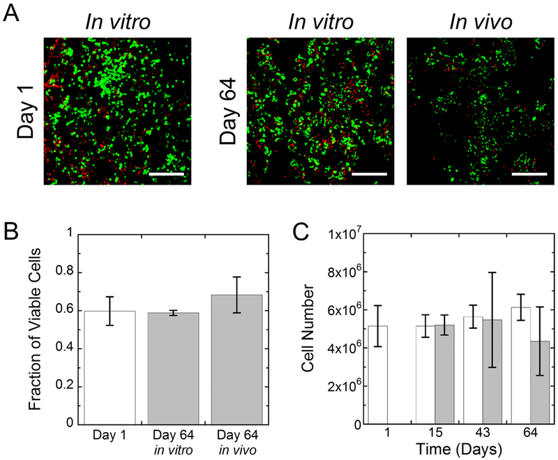
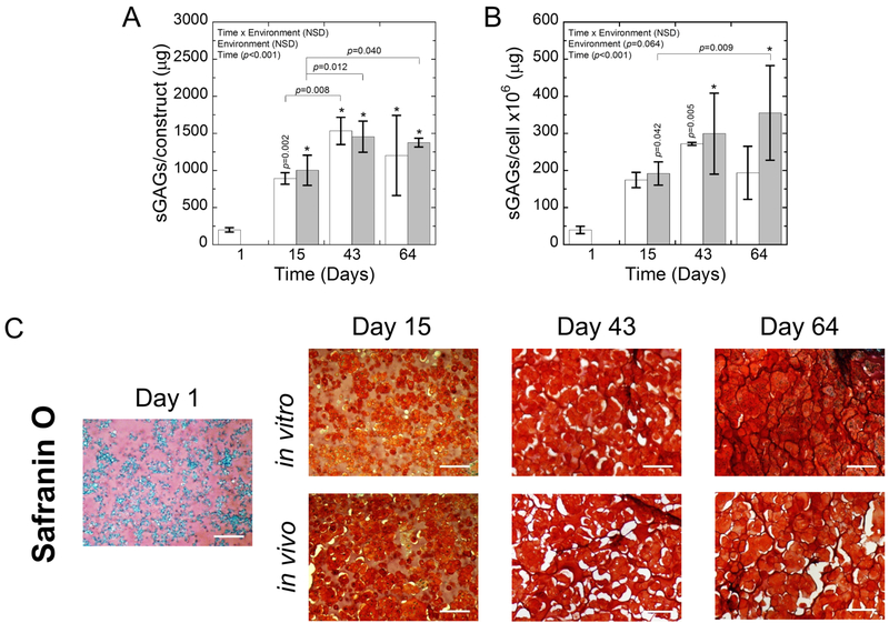
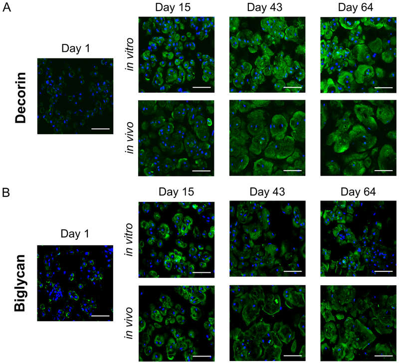
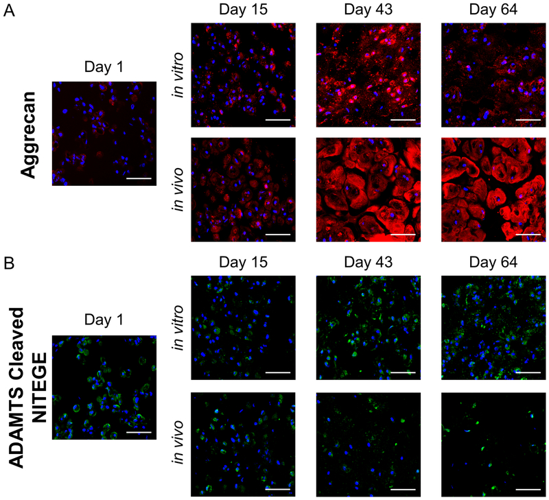
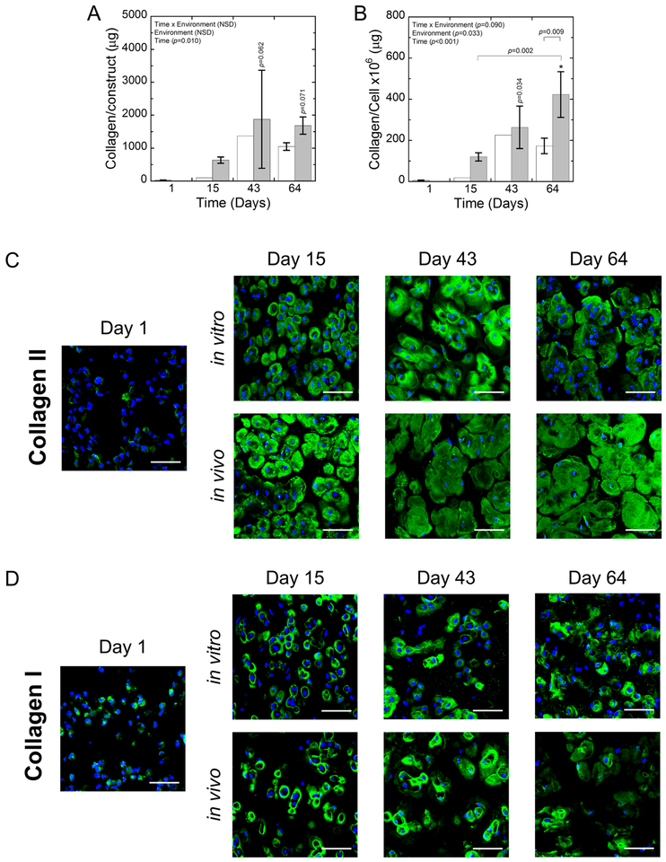
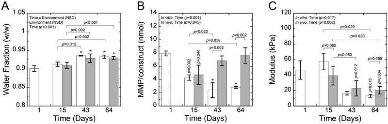
While matrix-assisted autologous chondrocyte implantation has emerged as a promising therapy to treat focal chondral defects, matrices that support regeneration of hyaline cartilage remain challenging. The goal of this work was to investigate the potential of a matrix metalloproteinase (MMP)-sensitive poly(ethylene glycol) (PEG) hydrogel containing the tethered growth factor, transforming growth factor β3 (TGF-β3), and compare cartilage regeneration in vitro and in vivo. The in vitro environment comprised chemically-defined medium while the in vivo environment utilized the subcutaneous implant model in athymic mice. Porcine chondrocytes were isolated and expanded in 2D culture for 10 days prior to encapsulation. The presence of tethered TGF-β3 reduced cell spreading. Chondrocyte-laden hydrogels were analyzed for total sulfated glycosaminoglycan and collagen contents, MMP activity, and spatial deposition of aggrecan, decorin, biglycan, and collagens type II and I. The total amount of extracellular matrix (ECM) deposited in the hydrogel constructs was similar in vitro and in vivo. However, the in vitro environment was not able to support long-term culture up to 64 days of the engineered cartilage leading to the eventual breakdown of aggrecan. The in vivo environment, on the other hand, led to more elaborate ECM, which correlated with higher MMP activity, and an overall higher quality of engineered tissue that was rich in aggrecan, decorin, biglycan and collagen type II with minimal collagen type I. Overall, the MMP-sensitive PEG hydrogel containing tethered TGF-β3 is a promising matrix for hyaline cartilage regeneration in vivo. STATEMENT OF SIGNIFICANCE: Regenerating hyaline cartilage remains a significant clinical challenge. The resultant repair tissue is often fibrocartilage, which long-term cannot be sustained. The goal of this study was to investigate the potential of a synthetic hydrogel matrix containing peptide crosslinks that can be degraded by enzymes secreted by encapsulated cartilage cells (i.e., chondrocytes) and tethered growth factors, specifically TGF-β3, to provide localized chondrogenic cues to the cells. This hydrogel led to hyaline cartilage-like tissue growth in vitro and in vivo, with minimal formation of fibrocartilage. However, the tissue formed in vitro, could not be maintained long-term. In vivo this hydrogel shows great promise as a potential matrix for use in regenerating hyaline cartilage.

Keywords: Cartilage extracellular matrix; Chondrocyte; In vitro; In vivo; Poly(ethylene glycol) hydrogel; Transforming growth factor beta 3.


Figures

Fig. 1:
Fig. 1:
A) Flow diagram showing the cartilage harvest, digestion, and 2D expansion of chondrocytes. B) The total cells recovered from the isolation and the number of cells recovered after 2D expansion.
Fig. 2:
Fig. 2:
A) Illustration of thiolated TGF-β3 tethering to PEG-NB via a photoclick reaction of thiol:norbornene. B) Illustration of PEG-NB-TGF crosslinked with an MMP-sensitive peptide in the presence of porcine chondrocytes with photoinitiator under UV light to form a 3D network. C) Experimental design for the in vitro and in vivo environments.
Fig. 3:
Fig. 3:
The absorbance measured from a modified ELISA quantifying the presence of TGF-β3 tethered into the network as a function of thiolated TGF-β3 concentration in the solution used to prepare the hydrogels. Data represent mean with standard deviation as error bars (n=3).
Fig. 4:
Fig. 4:
A) Representative confocal microscopy images of live cells (green) from days 1 and 29 for chondrocytes cultured in MMP-sensitive hydrogels without (−) or with (+) 50 nM tethered TGF-β3. Cell protrusions are noted with red arrowheads. Scale bar is 200 μm in low magnification images and 50 μm in high magnification images. B) Quantification of cell circularity at day 29. C) Quantification of cell roundness at day 29. B) Quantification of the fraction of cells with protrusions at day 29. –TGF-β3 constructs are shown in white. +TGF-β3 constructs are shown in gray. Data represent mean with standard deviation as error bars (n=2–3).
Fig. 5:
Fig. 5:
A) Representative confocal microscopy images of live (green) and dead (red) cells for in vitro and in vivo constructs at days 1 and 64. Scale bar is 200 μm. B) The fraction of viable cells based on live/dead images at day 1 (white bar) and for cells at day 64 (gray) for the in vitro and in vivo environments. C) The cell number for in vitro (white) and in vivo (gray) constructs. Data represent mean with standard deviation as error bars (n=3–6).
Fig. 6:
Fig. 6:
A) The sGAGs/construct and B) sGAGs/cell for in vitro (white) and in vivo (gray) constructs. Data represent mean with standard deviation as error bars. P-values shown vertically above a column indicate the difference from day 1. An * above a column indicates a statistical difference p<0.001 from day 1. C) Representative brightfield microscopy images of Safranin O stained sections for in vitro and in vivo constructs at days 1, 15, 43, and 64. sGAGs are stained red. Hematoxylin-stained nuclei are purple. The presence of white spaces in the images is the results of artifacts that arise during the dehydration and rehydration processes. Scale bar is 200 μm.
Fig. 7:
Fig. 7:
Representative confocal microscopy images for (A) decorin (green) and (B) biglycan (green) for in vitro and in vivo constructs at days 1, 15, 43, and 64. DAPI-stained nuclei are blue. Scale bar is 50 μm.
Fig. 8:
Fig. 8:
Representative confocal microscopy images for (A) aggrecan (red) and (B) the aggrecanase-cleaved epitope NITEGE (green) for in vitro and in vivo constructs at days 1, 15, 43, and 64. DAPI-stained nuclei are blue. Scale bar is 50 μm.
Fig. 9:
Fig. 9:
A) The collagen/construct and B) collagen/cell for in vitro (white) and in vivo (gray) constructs. Data represent mean with standard deviation as error bars. P-values shown vertically above a column indicate the difference from day 1. An * above a column indicates a statistical difference p<0.001 from day 1. B-C) Representative confocal microscopy images for (B) collagen II (green) and (C) collagen I (green) for in vitro and in vivo constructs at days 1, 15, 43, and 64. DAPI-stained nuclei are blue. Scale bar is 50 μm.
Fig. 10:
Fig. 10:
A) The water fraction, B) MMP activity/construct, and C) compressive modulus for in vitro (white) and in vivo (gray) constructs. Data represent mean with standard deviation as error bars. P-values shown vertically above a column indicate the difference from day 1. An * above a column indicates a statistical difference p<0.001 from day 1.

References

    1. Brittberg M, Recker D, Ilgenfritz J, Saris DBF, Matrix-Applied Characterized Autologous Cultured Chondrocytes Versus Microfracture: Five-Year Follow-up of a Prospective Randomized Trial, Am. J. Sports Med 46 (2018) 1343–1351. doi: 10.1177/0363546518756976. - DOI - PubMed
    1. Kon E, Delcogliano M, Filardo G, Montaperto C, Marcacci M, Second Generation Issues in Cartilage Repair, Sports Med. Arthrosc. Rev 16 (2008) 221–229. doi: 10.1097/JSA.0b013e31818cdbc5. - DOI - PubMed
    1. Schlegel W, Nuernberger S, Hombauer M, Albrecht C, Vecsei V, Marlovits S, Scaffold-dependent differentiation of human articular chondrocytes, Int. J. Mol. Med 22 (2008) 691–699. doi: 10.3892/ijmm_00000074. - DOI - PubMed
    1. Rowland CR, Lennon DP, Caplan AI, Guilak F, The Effects of Crosslinking of Scaffolds Engineered from Cartilage ECM on the Chondrogenic Differentiation of MSCs, Biomaterials. 34 (2013) 5802–5812. doi: 10.1016/j.biomaterials.2013.04.027. - DOI - PMC - PubMed
    1. Almeida HV, Liu Y, Cunniffe GM, Mulhall KJ, Matsiko A, Buckley CT, O’Brien FJ, Kelly DJ, Controlled release of transforming growth factor-β3 from cartilage-extra-cellular-matrix-derived scaffolds to promote chondrogenesis of human-joint-tissue-derived stem cells, Acta Biomater. 10 (2014) 4400–4409. doi: 10.1016/j.actbio.2014.05.030. - DOI - PubMed

Publication types

MeSH terms

LinkOut - more resources