. 2019 Mar 26;146(6):dev173146.
doi: 10.1242/dev.173146.

Defined conditions for propagation and manipulation of mouse embryonic stem cells

Affiliations

Defined conditions for propagation and manipulation of mouse embryonic stem cells

Carla Mulas et al. Development. .

Erratum in

Abstract

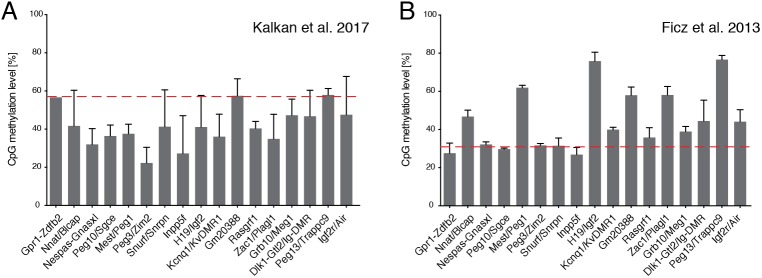
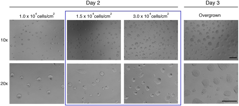
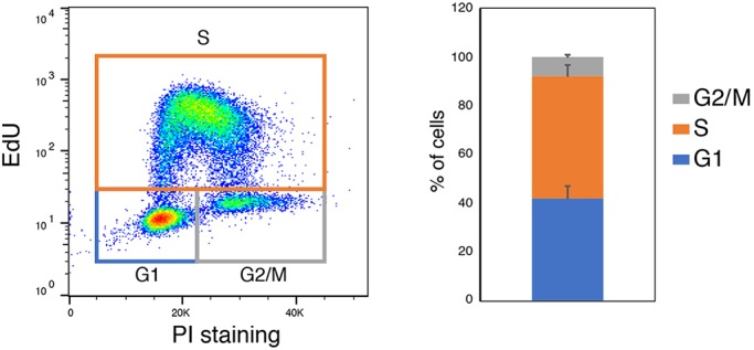
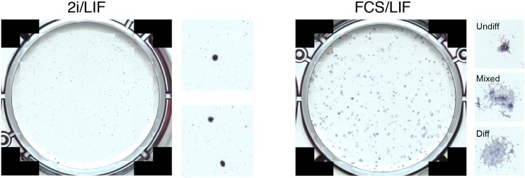
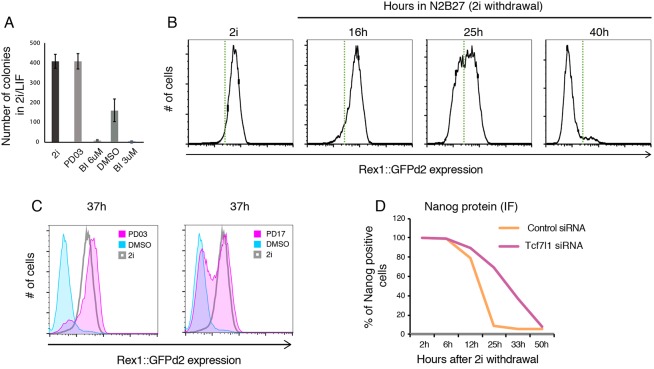
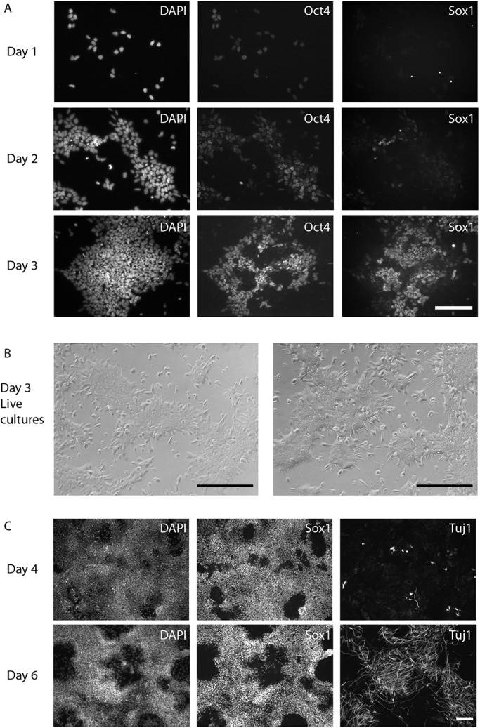
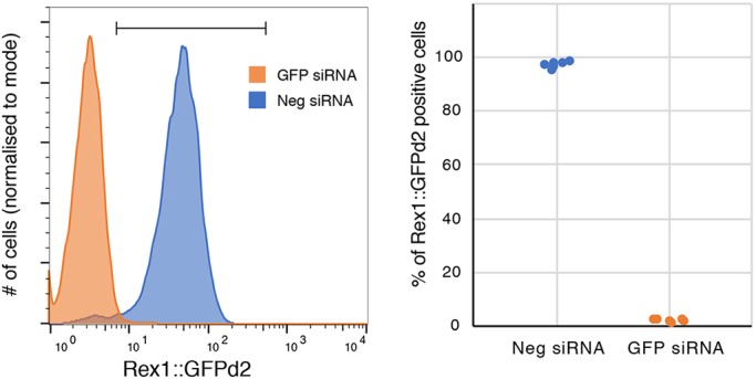
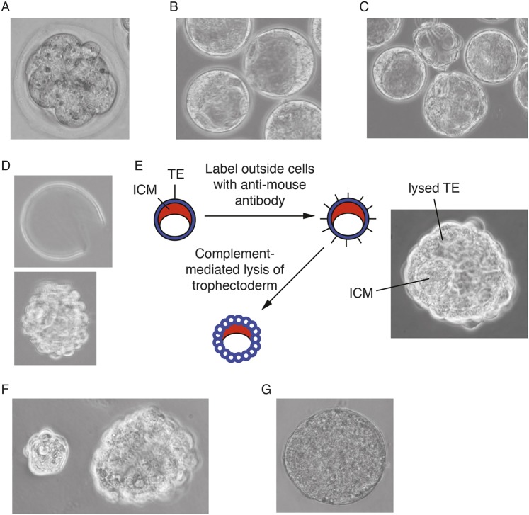
The power of mouse embryonic stem (ES) cells to colonise the developing embryo has revolutionised mammalian developmental genetics and stem cell research. This power is vulnerable, however, to the cell culture environment, deficiencies in which can lead to cellular heterogeneity, adaptive phenotypes, epigenetic aberrations and genetic abnormalities. Here, we provide detailed methodologies for derivation, propagation, genetic modification and primary differentiation of ES cells in 2i or 2i+LIF media without serum or undefined serum substitutes. Implemented diligently, these procedures minimise variability and deviation, thereby improving the efficiency, reproducibility and biological validity of ES cell experimentation.

Keywords: Differentiation; Embryonic stem cells; Pluripotency; Self-renewal.


Conflict of interest statement

Competing interestsThe authors declare no competing or financial interests.

Figures

Fig. 1.
Fig. 1.
Imprinted control region (ICR) methylation levels in mouse ESCs. (A,B) Average CpG methylation levels at known ICRs were quantified in whole genome bisulphite sequencing (WGBS) data sets from an ES cell line maintained in 2i (no LIF) (Kalkan et al., 2017; GEO accession number GSE92273) (A) and from an ES cell line maintained in 2i/LIF (Ficz et al., 2013; GEO accession number GSE42923) (B). Both datasets are derived from inbred 129 strain lines. The WGBS datasets were processed as described previously (von Meyenn et al., 2016) and the mean±s.d. of three experiments are shown. The mean global CpG methylation levels in each condition are shown (red dashed line). These observations are in agreement with previous reports that methylation at three DMRs is maintained in an ES cell line derived and maintained in 2i/LIF and an allele-specific assay confirmation of normal methylation pattern at the same regions in 2i/LIF-derived embryonic germ cells (Leitch et al., 2013a).
Fig. 2.
Fig. 2.
Representative images of ES cells in 2i at different densities on day 2 and day 3. Blue box highlights the range of cell densities ideal for splitting. Note refractile colony edges on day 2, which are lost in overgrown colonies at day 3. Scale bars: 0.5 mm.
Fig. 3.
Fig. 3.
Typical cell cycle profile of day 2 ES cells plated at 1.5×10⁴ cell/cm2. Cells were stained with propidium iodide (PI) and the Click-iT EdU kit according to manufacturer's instructions. Graph shows quantification over two independent experiments, with two separate lines in each.
Fig. 4.
Fig. 4.
Representative images of clonal assays in 2i/LIF and FCS/LIF conditions. Insets to the right show magnified views of individual colonies and suggested classification in the case of FCS/LIF.
Fig. 5.
Fig. 5.
Example data of exit from pluripotency experiment. (A) Colony-formation assay. Cells were differentiated for 36 h in four different conditions; PD03, MEK1/2 inhibitor PD0325901 (1 µM); BI, RSK inhibitor BI-D1870 (3 or 6 µM); DMSO, carrier control. (B) Representative downregulation kinetics of Rex1::GFPd2 cells plated in N2B27, starting from 2i (no LIF) conditions. Green dotted line indicates arbitrary threshold. (C) Use of the Rex1::GFPd2 reporter system to determine the effect of two different inhibitors (PD03, MEK1/2 inhibitor PD0325901 at 1 µM; PD17, FGF receptor inhibitor PD173074 at 100 nM) on exit kinetics. (D) Fixing and immunostaining for Nanog protein, to quantify the delay in transition associated with knockdown of the Tcf7l1 transcription factor by siRNA.
Fig. 6.
Fig. 6.
Representative images of cells at different stages of neural differentiation. (A) Immunostaining for Sox1 and Oct4 on day 1-3 of neural differentiation. (B) Phase contrast images showing representative morphology of early day 3 neural differentiation for two different cell lines (live cultures). Little cell death should occur during the first 1-3 days of differentiation. Cell death might become apparent once cultures become confluent. (C) Immunostaining for Sox1 and the post-mitotic marker Tuj1 on days 4 and 6 of differentiation. Scale bars: 0.5 mm.
Fig. 7.
Fig. 7.
Transfection efficiency as assessed by knockdown of GFP in Rex1::GFPd2 cells. Left: representative flow cytometry profile. Right: quantification of the percentage of Rex1::GFPd2 positive cells over four independent experiments.
Fig. 8.
Fig. 8.
ES cell derivation process in 2i/LIF. (A) Flushed 8-cell embryo. (B) Embryos after 1-2 days in culture. (C) After 3 days in culture, embryos start to hatch from the zona pellucida. (D) Hatched blastocyst (bottom) and discarded zona (top). (E) Summary of immunosurgery protocol and image of ideal end-point after complement treatment. (F) Separation of ICM and trophectoderm after immunosurgery. (G) Primary outgrowth cultured for 4 days in 2i/LIF on gelatinised plates (not to scale with embryos). Images reproduced with permission from Cold Spring Harbor Laboratory Press. These images are not published under the terms of the CC-BY licence of this article. For permission to reuse, please see Nichols and Jones, 2017. TE, trophectoderm.

References

    1. Andersson-Rolf A., Mustata R. C., Merenda A., Kim J., Perera S., Grego T., Andrews K., Tremble K., Silva J. C. R., Fink J. et al. (2017). One-step generation of conditional and reversible gene knockouts. Nat. Methods 14, 287-289. 10.1038/nmeth.4156 - DOI - PMC - PubMed
    1. Batlle-Morera L., Smith A. and Nichols J. (2008). Parameters influencing derivation of embryonic stem cells from murine embryos. Genesis 46, 758-767. 10.1002/dvg.20442 - DOI - PubMed
    1. Betschinger J., Nichols J., Dietmann S., Corrin P. D., Paddison P. J. and Smith A. (2013). Exit from pluripotency is gated by intracellular redistribution of the bHLH transcription factor Tfe3. Cell 153, 335-347. 10.1016/j.cell.2013.03.012 - DOI - PMC - PubMed
    1. Blaschke K., Ebata K. T., Karimi M. M., Zepeda-Martínez J. A., Goyal P., Mahapatra S., Tam A., Laird D. J., Hirst M., Rao A. et al. (2013). Vitamin C induces Tet-dependent DNA demethylation and a blastocyst-like state in ES cells. Nature 500, 222-226. 10.1038/nature12362 - DOI - PMC - PubMed
    1. Boroviak T., Loos R., Bertone P., Smith A. and Nichols J. (2014). The ability of inner-cell-mass cells to self-renew as embryonic stem cells is acquired following epiblast specification. Nat. Cell Biol. 16, 516-528. 10.1038/ncb2965 - DOI - PMC - PubMed

Publication types