

doi: 10.1182/blood-2019-01-895698.
Epub 2019 Mar 26.
Familial predisposition to TP53/complex karyotype MDS and leukemia in DNA repair-deficient xeroderma pigmentosum
Affiliations
- PMID: 30914417
- PMCID: PMC6610036
- DOI: 10.1182/blood-2019-01-895698
Item in Clipboard
Familial predisposition to TP53/complex karyotype MDS and leukemia in DNA repair-deficient xeroderma pigmentosum
Blood.
.
Abstract
There is a
Conflict of interest statement
Conflict-of-interest disclosure: The authors declare no competing financial interests.
Figures
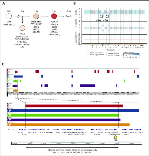
Germline genetics and clonal evolution in patients with XP-C with MDS and leukemia. (A) Bone marrow progression in patient #5 (XP924VI) showing clonal evolution. Chromosomal abnormalities and somatic point mutations are shown. (B) Copy number and allele heterozygosity analysis of the bone marrow MDS-EB1 sample showing deletion 5q and 7q (arrows) in patient #5. WES data were analyzed using the FACETS tool; total copy number log-ratio (logR; upper); allele-specific log-odds-ratio data (logOR; middle); corresponding integer (total, minor) copy number calls (bottom). The estimated cellular fraction (cf) profile is plotted at the bottom. (C) Minimal common region of germinal homozygosity in 3p25 including the XPC gene (yellow arrow), as shown by the analysis of single nucleotide polymorphism array data in the 5 patients with available fibroblast cell DNA (Affymetrix array) and confirmed using WES data (Illumina). Data are shown using the Genome Wide SNP6 Array and the Chromosome Analysis Suite. Loss of heterozygosity segments on chromosome 3 (top); minimal region of homozygosity as mapped by patient XP10VI and XP309VI, on the left and right sides (genomic position on chr3: 11 092 104 and 14 285 238, using hg19 reference, NM_00628.4), respectively (bottom).
Comment in
-
FouNdER mutations confer risk for leukemias.Blood. 2019 Jun 20;133(25):2636-2638. doi: 10.1182/blood-2019-04-901173. Blood. 2019. PMID: 31221794 No abstract available.
References
-
- Kraemer KH, Sarasin A. Xeroderma pigmentosum. In: Elder DE, Massi D, Scolyer RA, Willemze R, eds. WHO Classification of Skin Tumours, 4th ed Lyon, France: IARC Press; 2018:386-387.
-
- Hanawalt PC, Spivak G. Transcription-coupled DNA repair: two decades of progress and surprises. Nat Rev Mol Cell Biol. 2008;9(12):958-970. - PubMed
-
- Soufir N, Ged C, Bourillon A, et al. . A prevalent mutation with founder effect in xeroderma pigmentosum group C from north Africa. J Invest Dermatol. 2010;130(6):1537-1542. - PubMed
-
- Desandes E, Lacour B, Belot A, et al. . Cancer incidence and survival in adolescents and young adults in France, 2000-2008. Pediatr Hematol Oncol. 2013;30(4):291-306. - PubMed
Publication types
MeSH terms
Substances
Supplementary concepts
LinkOut - more resources
Full Text Sources
Medical
Research Materials
Miscellaneous
