. 2019 Dec;68(12):2142-2151.
doi: 10.1136/gutjnl-2018-317571. Epub 2019 Mar 26.

Structural weakening of the colonic mucus barrier is an early event in ulcerative colitis pathogenesis

Affiliations

Structural weakening of the colonic mucus barrier is an early event in ulcerative colitis pathogenesis

Sjoerd van der Post et al. Gut. 2019 Dec.

Abstract

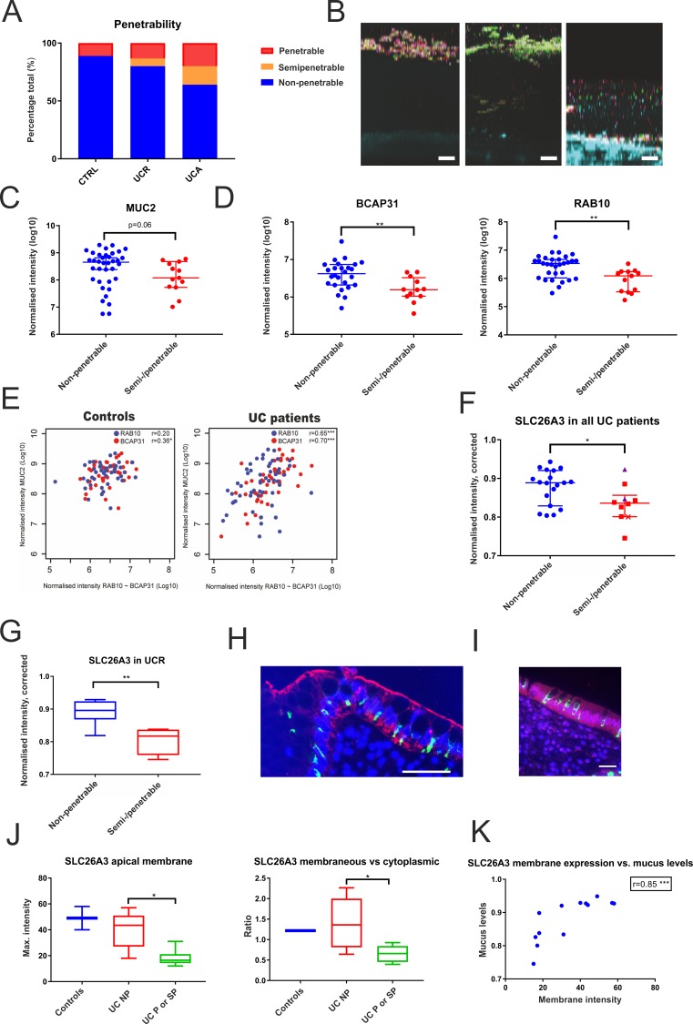
Objective: The colonic inner mucus layer protects us from pathogens and commensal-induced inflammation, and has been shown to be defective in active UC. The aim of this study was to determine the underlying compositional alterations, their molecular background and potential contribution to UC pathogenesis.

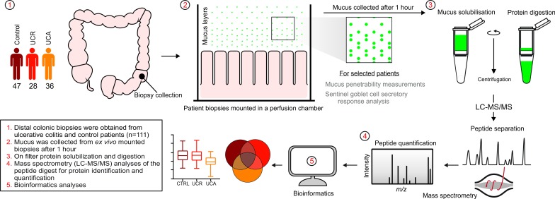
Design: In this single-centre case-control study, sigmoid colon biopsies were obtained from patients with UC with ongoing inflammation (n=36) or in remission (n=28), and from 47 patients without colonic disease. Mucus samples were collected from biopsies ex vivo, and their protein composition analysed by nanoliquid chromatography-tandem mass spectrometry. Mucus penetrability and goblet cell responses to microbial stimulus were assessed in a subset of patients.

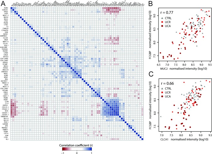
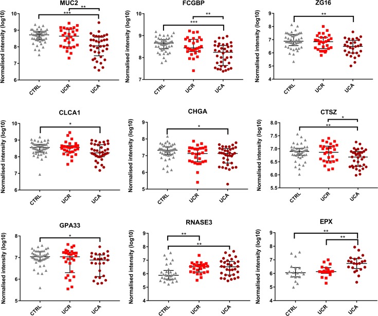
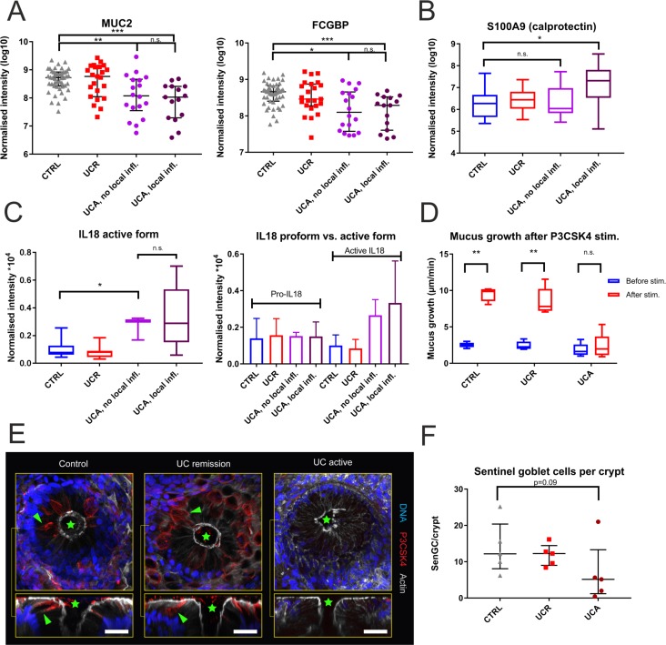
Results: The core mucus proteome was found to consist of a small set of 29 secreted/transmembrane proteins. In active UC, major structural mucus components including the mucin MUC2 (p<0.0001) were reduced, also in non-inflamed segments. Active UC was associated with decreased numbers of sentinel goblet cells and attenuation of the goblet cell secretory response to microbial challenge. Abnormal penetrability of the inner mucus layer was observed in a subset of patients with UC (12/40; 30%). Proteomic alterations in penetrable mucus samples included a reduction of the SLC26A3 apical membrane anion exchanger, which supplies bicarbonate required for colonic mucin barrier formation.

Conclusion: Core mucus structural components were reduced in active UC. These alterations were associated with attenuation of the goblet cell secretory response to microbial challenge, but occurred independent of local inflammation. Thus, mucus abnormalities are likely to contribute to UC pathogenesis.

Keywords: mucins; mucosal barrier; mucosal protection; mucus; ulcerative colitis.


Conflict of interest statement

Competing interests: None declared.

Figures

Figure 1
Figure 1
Schematic overview of the study design and workflow of the mass spectrometry analysis of colonic mucus samples.  LC-MS/MS, liquid chromatography-tandem mass spectrometry; m/z, mass-to-charge ratio; UC, ulcerative colitis.
Figure 2
Figure 2
Correlations between the mucus proteins. (A) Matrix of Pearson r correlation coefficients for all members of the mucus proteome (71 proteins), based on their normalised intensities in all patient samples (n=111). Correlation of normalised intensities of (B) MUC2 and FCGBP (r=0.77; p<0.0001) and (C) FCGBP and CLCA1 (r=0.66; p<0.0001). Patients with active UC (UCA) and UC in remission (UCR) are denoted as circles and squares, respectively; controls (CTRL) as triangles.
Figure 3
Figure 3
Core mucus proteins that were significantly altered in active UC. Data for the individual patients are overlaid by the median and IQR. Significance was determined by one-way analyses of variance (ANOVA) with Tukey’s post hoc test, followed by pairwise Mann-Whitney U tests (*p≤0.05; **p≤0.01; ***p≤0.001). CTRL, controls; UCA, active UC; UCR, UC in remission.
Figure 4
Figure 4
The reduction in mucus core proteins is not directly caused by inflammation, but likely related to exhaustion of the upper crypt goblet cell secretory response to microbes. (A) FCGBP and MUC2 levels did not differ between patients with clinically active UC with and without inflammation of the sigmoid colon. Data are overlaid by the median and IQR. Inflammation was defined as a Sandborn score ≥2. (B) Mucus levels of the neutrophil protein S100A9, part of the calprotectin dimer, were not increased in patients with clinically active UC without local inflammation. (C) Mucus levels of the active part of IL18 were equally increased in patients with clinically active UC with and without local inflammation. Quantification based on two unique peptides (SDIIFFQR and SIMFTVQNED.-). The levels of the inactive IL18 propeptide did not differ between patient groups, based on one unique peptide (AAEPVEDNCINFVAMK). Bars represent the mean; error bars 1 SD. One control that was diagnosed with colon cancer during follow-up was excluded from analysis. (D) Sentinel goblet cell secretory response to the bacterial Toll-like receptor (TLR) 1/2-ligand P3CSK4 was abrogated in active UC. The box plot shows data from five controls, five patients with UC in remission and five patients with active UC. (E) The number of sentinel goblet cells (red) was reduced in patients with active UC. The upper panel shows cross sections (x/y axis view) of a representative sigmoid colon biopsy from each patient category; the lower panel longitudinal (x/z axis) views. Green stars represent crypt openings; green arrowheads represent examples of sentinel goblet cells. Sentinel goblet cells were identified as upper crypt goblet cells that had endocytosed fluorescently labelled P3CSK4. Scale bars: 30 µm. (F) Mean number of sentinel goblet cells per crypt for each patient category. Individual observations are overlaid with median and IQR. The high outlier among the patients with active UC had commenced peroral cortisone treatment 3 weeks prior to colonoscopy. For all box plots, the line represents the median, the box the IQR and the whiskers the range. Groups were compared by the Mann-Whitney U test with Bonferroni correction for multiple comparisons. CTRL, controls; IL18, interleukin 18; n.s., not significant; UCA, active UC; UCR, UC in remission.
Figure 5
Figure 5
Changes in mucus protein composition associated with inner mucus layer failure. (A) Proportions of penetrable, semipenetrable and non-penetrable mucus samples among patients with UC and controls. (B) Representative confocal images of (from left to right) impenetrable, semipenetrable and penetrable mucus. The blue colour represents the epithelium; red, purple and green dots represent fluorescent beads with diameters of 0.5, 1 and 2 µm, respectively. (C) MUC2 levels in non-penetrable and semi/penetrable mucus. (D) Levels of putative compound exocytosis markers BCAP31 and RAB10 in non-penetrable and semi/penetrable mucus. (E) Correlations of normalised intensities between MUC2 levels and BCAP31 and RAB10, respectively, for patients with UC and for the control group. R values refer to the Spearman’s rank correlation coefficient. (F) Levels of chloride-bicarbonate transporter SLC26A3 (DRA) were reduced in semi/penetrable mucus from patients with UC. Triangles represent two patients with active disease who regained an impenetrable inner mucus layer in remission, whereas the x represents a patient in remission, whose mucus remained penetrable upon reanalysis after 3 years. (G) The reduction in mucus SLC26A3 was particularly pronounced in individuals in remission. One borderline case with clinical Mayo score 2, endoscopic Mayo score 1 and Sandborn score 1 was included among the remission patients. In (F) and (G), SLC26A3 abundance factors were corrected for the degree of mucus cellular contamination through the division of SLC26A3 normalised intensity values by the median normalised intensity of the top 100 most abundant intracellular proteins, for each sample. (H) SLC26A3 (red) was apically localised in both colonocytes and surface goblet cells. Costaining of non-O-glycosylated apomucin 2 (localised to the endoplasmic reticulum [ER]; green) identifies goblet cells; nuclei are stained blue. Scale bar: 50 μm. (I) In patients with UC with penetrable mucus, membrane staining for SLC26A3 (red) appeared weaker. Scale bar: 25 µm. (J) The left panel shows a comparison of the intensity of SLC26A3 apical membrane staining for patients with UC with and without penetrable mucus. The right panel shows the ratio between membrane and cytoplasmic SLC26A3 staining intensity for each patient category. Results are based on immunohistochemical analysis of patients with UC with (semi)penetrable mucus (n=6), patients with UC with non-penetrable mucus (n=6) and controls (n=3), and represent averages of 15 measurements. (K) Correlation between SLC26A3 apical membrane staining intensity and SLC26A3 mucus levels according to relative quantification by proteomics. The r value refers to the Spearman’s rank correlation coefficient. For all box plots, the line represents the median, the box the IQR and the whiskers the range of values. Groups were compared by the Mann-Whitney U test. *p≤0.05; **p≤0.01; ***p≤0.001. CTRL, controls; NP, non-penetrable; SP, semipenetrable; P, penetrable; UCA, active UC; UCR, UC in remission.

References

    1. Danese S, Fiocchi C. Ulcerative colitis. N Engl J Med 2011;365:1713–25. 10.1056/NEJMra1102942 - DOI - PubMed
    1. Johansson ME, Phillipson M, Petersson J, et al. . The inner of the two Muc2 mucin-dependent mucus layers in colon is devoid of bacteria. Proc Natl Acad Sci U S A 2008;105:15064–9. 10.1073/pnas.0803124105 - DOI - PMC - PubMed
    1. Van der Sluis M, De Koning BA, De Bruijn AC, et al. . Muc2-deficient mice spontaneously develop colitis, indicating that MUC2 is critical for colonic protection. Gastroenterology 2006;131:117–29. 10.1053/j.gastro.2006.04.020 - DOI - PubMed
    1. Velcich A, Yang W, Heyer J, et al. . Colorectal cancer in mice genetically deficient in the mucin Muc2. Science 2002;295:1726–9. 10.1126/science.1069094 - DOI - PubMed
    1. Blumberg RS, Saubermann LJ, Strober W. Animal models of mucosal inflammation and their relation to human inflammatory bowel disease. Curr Opin Immunol 1999;11:648–56. 10.1016/S0952-7915(99)00032-1 - DOI - PubMed

Publication types

MeSH terms