. 2019 Apr 9;116(15):7288-7297.
doi: 10.1073/pnas.1815027116. Epub 2019 Mar 26.

Crystal structure and activity-based labeling reveal the mechanisms for linkage-specific substrate recognition by deubiquitinase USP9X

Affiliations

Crystal structure and activity-based labeling reveal the mechanisms for linkage-specific substrate recognition by deubiquitinase USP9X

Prajwal Paudel et al. Proc Natl Acad Sci U S A. .

Abstract

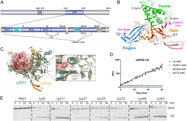
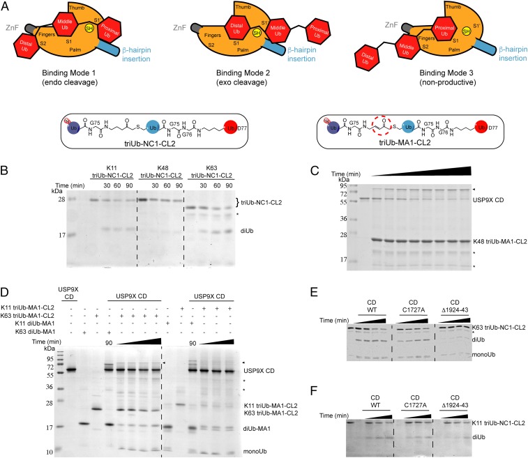
USP9X is a conserved deubiquitinase (DUB) that regulates multiple cellular processes. Dysregulation of USP9X has been linked to cancers and X-linked intellectual disability. Here, we report the crystal structure of the USP9X catalytic domain at 2.5-Å resolution. The structure reveals a canonical USP-fold comprised of fingers, palm, and thumb subdomains, as well as an unusual β-hairpin insertion. The catalytic triad of USP9X is aligned in an active configuration. USP9X is exclusively active against ubiquitin (Ub) but not Ub-like modifiers. Cleavage assays with di-, tri-, and tetraUb chains show that the USP9X catalytic domain has a clear preference for K11-, followed by K63-, K48-, and K6-linked polyUb chains. Using a set of activity-based diUb and triUb probes (ABPs), we demonstrate that the USP9X catalytic domain has an exo-cleavage preference for K48- and endo-cleavage preference for K11-linked polyUb chains. The structure model and biochemical data suggest that the USP9X catalytic domain harbors three Ub binding sites, and a zinc finger in the fingers subdomain and the β-hairpin insertion both play important roles in polyUb chain processing and linkage specificity. Furthermore, unexpected labeling of a secondary, noncatalytic cysteine located on a blocking loop adjacent to the catalytic site by K11-diUb ABP implicates a previously unreported mechanism of polyUb chain recognition. The structural features of USP9X revealed in our study are critical for understanding its DUB activity. The new Ub-based ABPs form a set of valuable tools to understand polyUb chain processing by the cysteine protease class of DUBs.

Keywords: USP9X; activity-based probes; deubiquitinase; linkage specificity; zinc finger.


Conflict of interest statement

The authors declare no conflict of interest.

Figures

Fig. 1.
Fig. 1.
Structure and activity of USP9X. (A) Domain architecture of the full-length USP9X and the CD. Sites for surface entropy reduction mutations (K1637E1638) and ZnF mutations (Cys1727 or H1729) are marked with an asterisk (*) and pound sign (#), respectively. Blocking loops are colored yellow. (B) Crystal structure of the USP9X CD. The fingers, palm, and thumb subdomains are colored blue, orange, and green, respectively. The β-hairpin insertion (Ins3) is salmon-colored, and the two blocking loops are colored in red. Residues in the catalytic triad and the ZnF are shown in sticks. The zinc ion is shown as a sphere. S2, S1, and S1′ indicate three potential Ub binding sites. (C) Superposition of the USP9X and USP7/Ub-aldehyde (PDB ID code 1NBF) structures. Ub-aldehyde is shown in surface and ribbon representations (red), whereas USP7 (teal) and USP9X (orange) are shown in ribbon. (Right) A zoomed-in view of the overlay of the active sites of USP7 and USP9X. The catalytic triad residues in USP9X and USP7 and Ub Gly76 are labeled. (D) Activity assay using fluorogenic substrates showing that USP9X CD hydrolyzes Ub-AMC substrate efficiently but not SUMO1-, NEDD8-, or ISG15-AMC. (E) Gel-based cleavage assay of the USP9X CD using native M1-, K6-, K11-, K27-, K29-, K33-, K48-, and K63-diUb substrates.
Fig. 2.
Fig. 2.
Activity-based di- and triUb probes. (A) Structures and synthesis of the diUb-NC1-PA2 probes and diUb-NC1-AMC2 fluorogenic substrates. (B) Generation of two types of hybrid triUb probes using a chemoenzymatic approach. TriUb-NC1-CL2 contains a noncleavable bond between the distal (light purple) and middle (blue) Ubs and a native isopeptide linkage between the middle and proximal (red) Ubs. TriUb-MA1-CL2 contains an MA warhead between the distal and middle Ubs and a native isopeptide linkage between the middle and proximal Ubs.
Fig. 3.
Fig. 3.
Interrogation of the role of β-hairpin in proximal Ub recognition (A) Superposition of the USP9X CD apo structure with CYLD in complex with M1-diUb (PDB ID code 3WXE). USP9X, CYLD, and diUb are colored orange, purple, and red, respectively. The CYLD and USP9X insertions are circled and labeled. (B) Cleavage activity of WT USP9X CD and deletion mutant Δ(1924–1943) assayed using IQF-diUb substrates of K11 and K63 linkages. (C) Labeling of WT and β-hairpin deletion mutant USP9X CD by K11 diUb-MA1 probe at different time points.
Fig. 4.
Fig. 4.
A noncatalytic Cys1808 in USP9X CD is labeled by the K11-diUb-MA1 probe. (A) Labeling band pattern of the WT and mutant USP9X CD by K11 diUb-MA1 probe. C1566S is a catalytically dead mutant but is labeled at C1808 position. Mutation of the noncatalytic cysteine Cys1808 to Ser abolished the HMW labeling band but retained the LMW labeling band. (B) HA-Ub-VME only labels the catalytic Cys1566 but not Cys1808 and is not affected by ZnF mutation. (C) Surface representation of USP9X CD revealing a groove between the blocking loops BL1 (orange) and BL2 (green). Noncatalytic Cys1808 (yellow) labeled by K11-diUb-MA1 is on BL1 facing the groove.
Fig. 5.
Fig. 5.
USP9X CD exhibits linkage-specific endo/exo recognition of polyUb chains. (A) Illustration of the three potential binding modes of a triUb to the S2, S1, and S1′ binding sites of USP9X CD. (B) Cleavage of the K11-, K48-, and K63-linked triUb-NC1-CL2 by WT USP9X CD at three different time points. (C) Incubation of WT USP9X CD with K48 triUb-MA1-CL2 probe at 0-, 5-, 10-, 15-, 30-, 45-, 60-, and 90-min time points. (D) Labeling of WT USP9X CD by K63- and K11-linked triUb-MA1-CL2 probes at 15-, 30-, 60-, and 90-min time points and 30-, 60-, and 90-min time points, respectively. Labeling of WT USP9X CD by K63- and K11-linked diUb-MA1 probes is included for comparison. (E) Comparison of cleavage of K63- and K11- (F) triUb-NC1-CL2 by USP9X WT, ZnF mutant (C1727A) and deletion mutant (Δ1924–43) up to 120 min and 90 min, respectively. The reaction products were analyzed by SDS/PAGE and Coomassie brilliant blue staining. The asterisk denotes contaminating band in the probe preparations and triangle denotes labeling band.

References

    1. Nijman SM, et al. A genomic and functional inventory of deubiquitinating enzymes. Cell. 2005;123:773–786. - PubMed
    1. Ye Y, Scheel H, Hofmann K, Komander D. Dissection of USP catalytic domains reveals five common insertion points. Mol Biosyst. 2009;5:1797–1808. - PubMed
    1. Komander D, Clague MJ, Urbé S. Breaking the chains: Structure and function of the deubiquitinases. Nat Rev Mol Cell Biol. 2009;10:550–563. - PubMed
    1. Faesen AC, et al. The differential modulation of USP activity by internal regulatory domains, interactors and eight ubiquitin chain types. Chem Biol. 2011;18:1550–1561. - PubMed
    1. Hu M, et al. Crystal structure of a UBP-family deubiquitinating enzyme in isolation and in complex with ubiquitin aldehyde. Cell. 2002;111:1041–1054. - PubMed

Publication types

Associated data

LinkOut - more resources