. 2019 Mar 26;10(2):e00319-19.
doi: 10.1128/mBio.00319-19.

Antagonism of dsRNA-Induced Innate Immune Pathways by NS4a and NS4b Accessory Proteins during MERS Coronavirus Infection

Affiliations

Antagonism of dsRNA-Induced Innate Immune Pathways by NS4a and NS4b Accessory Proteins during MERS Coronavirus Infection

Courtney E Comar et al. mBio. .

Abstract

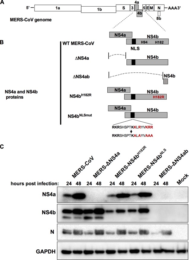
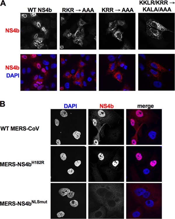
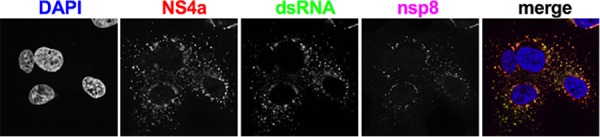
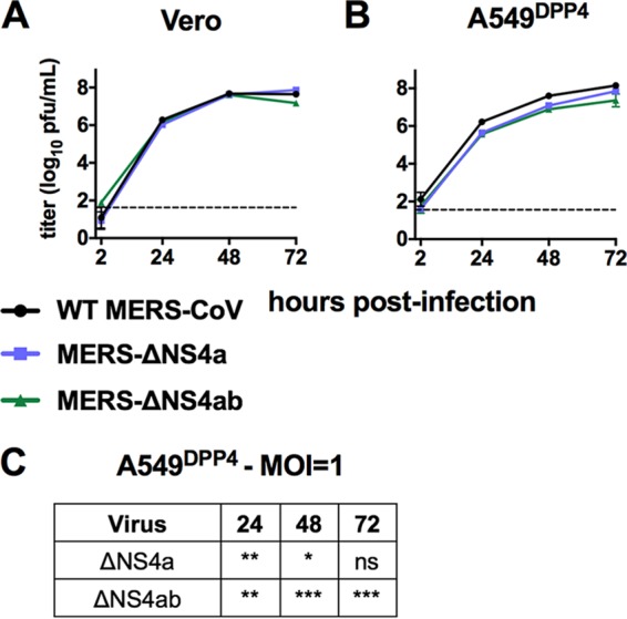
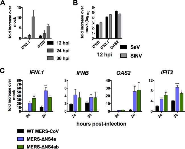
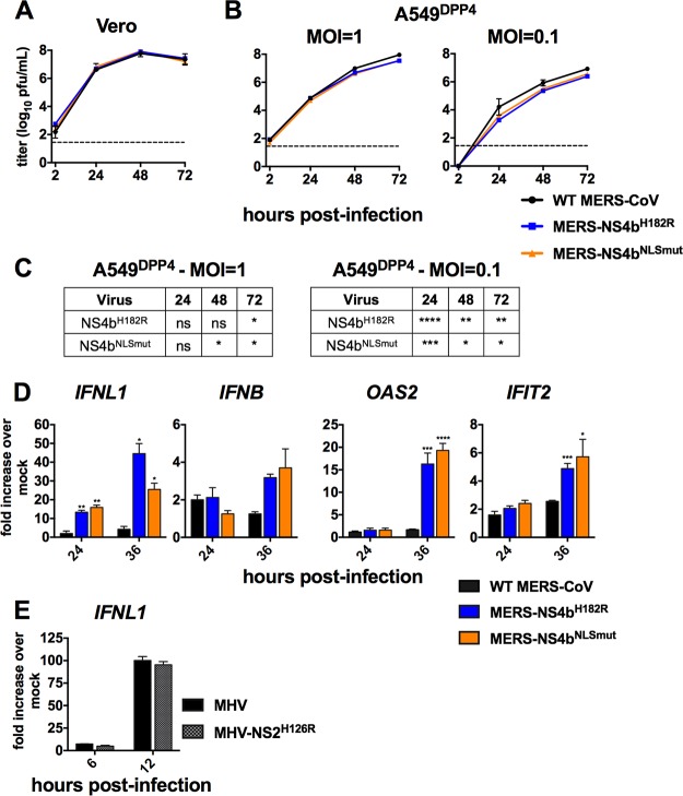
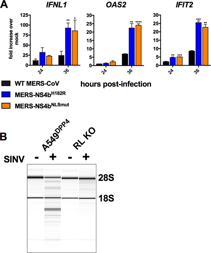
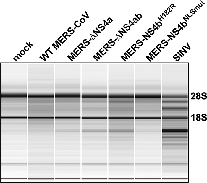
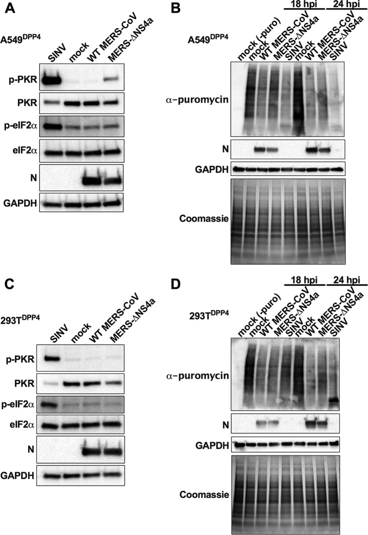
Middle East respiratory syndrome coronavirus (MERS-CoV) was first identified in 2012 as a novel etiological agent of severe respiratory disease in humans. As during infection by other viruses, host sensing of viral double-stranded RNA (dsRNA) induces several antiviral pathways. These include interferon (IFN), oligoadenylate synthetase (OAS)-RNase L, and protein kinase R (PKR). Coronaviruses, including MERS-CoV, potently suppress the activation of these pathways, inducing only modest host responses. Our study describes the functions of two accessory proteins unique to MERS-CoV and related viruses, NS4a and NS4b, during infection in human airway epithelium-derived A549 cells. NS4a has been previously characterized as a dsRNA binding protein, while NS4b is a 2',5'-phosphodiesterase with structural and enzymatic similarity to NS2 encoded by mouse hepatitis virus (MHV). We found that deletion of NS4a results in increased interferon lambda (IFNL1) expression, as does mutation of either the catalytic site or nuclear localization sequence of NS4b. All of the mutant viruses we tested exhibited slight decreases in replication. We previously reported that, like MHV NS2, NS4b antagonizes OAS-RNase L, but suppression of IFN is a previously unidentified function for viral phosphodiesterases. Unexpectedly, deletion of NS4a does not result in robust activation of the PKR or OAS-RNase L pathways. Therefore, MERS-CoV likely encodes other proteins that contribute to suppression or evasion of these antiviral innate immune pathways that should be an important focus of future work. This study provides additional insight into the complex interactions between MERS-CoV and the host immune response.IMPORTANCE Middle East respiratory syndrome coronavirus (MERS-CoV) is the second novel zoonotic coronavirus to emerge in the 21st century and cause outbreaks of severe respiratory disease. More than 2,200 cases and 800 deaths have been reported to date, yet there are no licensed vaccines or treatments. Coronaviruses encode unique accessory proteins that are not required for replication but most likely play roles in immune antagonism and/or pathogenesis. Our study describes the functions of MERS-CoV accessory proteins NS4a and NS4b during infection of a human airway-derived cell line. Loss of these accessory proteins during MERS-CoV infection leads to host antiviral activation and modestly attenuates replication. In the case of both NS4a and NS4b, we have identified roles during infection not previously described, yet the lack of robust activation suggests much remains to be learned about the interactions between MERS-CoV and the infected host.

Keywords: MERS-CoV; coronavirus; interferon antagonist; viral accessory proteins.


Figures

FIG 1
FIG 1
MERS-CoV NS4a and NS4b recombinant mutants. (A) MERS-CoV genome RNA with open reading frames shown. (B) NS4a and NS4b proteins expressed by wild-type and mutant MERS-CoVs. The catalytic His residues of the PDE are shown, and the vertical black bar indicates the NLS of NS4b; the red lettering indicates amino acid substitutions of the catalytic His residue and within the NLS. (C) Expression of viral proteins from recombinant MERS-CoV viruses. A549DPP4 cells were infected at an MOI of 10 with WT MERS-CoV, MERS-ΔNS4a, MERS-ΔNS4ab, MERS-NS4bH182R, or MERS-NS4bNLSmut or mock infected. Cell lysates were prepared at 24 and 48 h postinfection, analyzed by SDS-PAGE, and probed by Western blotting with rabbit antiserum against NS4a and NS4b or mouse monoclonal antibodies against MERS nucleocapsid protein (N) and GAPDH. The Western blot data are from one representative of three independent infections.
FIG 2
FIG 2
Subcellular localization of MERS-CoV NS4b expression. (A) The nuclear localization signal (NLS) was mapped by mutating basic residues in pCAGGS-NS4b, and NS4b was ectopically expressed in A549 cells by DNA transfection. Twenty-four hours posttransfection, cells were fixed and stained for NS4b using anti-NS4b rabbit serum and goat anti-rabbit AF594 secondary antibody. (B) A549DPP4 cells were infected with WT MERS-CoV, MERS-NS4bH182R, or MERS-NS4bNLSmut (MOI = 5). Cells were fixed 24 h postinfection and stained with anti-NS4b rabbit serum and goat anti-rabbit AF594 secondary antibody. The images shown in both panels are representative of at least three fields of cells from three independent experiments.
FIG 3
FIG 3
NS4a colocalizes with dsRNA around replication/transcription complexes (RTC) during MERS-CoV infection. A549DPP4 cells were infected with WT MERS-CoV (MOI = 5), fixed 24 h postinfection, and stained with rabbit anti-NS4a serum, mouse anti-dsRNA J2, and guinea pig anti-nsp8 serum and then with secondary antibodies goat anti-rabbit AF647, goat anti-mouse AF488, and goat anti-guinea pig AF568. The images shown are representative of at least three fields of cells from three independent experiments.
FIG 4
FIG 4
MERS-CoV NS4a and NS4b mutants are attenuated in IFN competent cells. (A) Vero cells were infected in triplicate at an MOI of 1 with WT MERS-CoV, MERS-ΔNS4a, and MERS-ΔNS4ab. Supernatants were collected at indicated times postinfection, and infectious virus was quantified by plaque assay. (B) A549DPP4 cells were infected in triplicate at an MOI of 1 with WT MERS-CoV, MERS-ΔNS4a, and MERS-ΔNS4ab, and replication was quantified as in panel A. (C) Statistical significance for mutant virus replication versus WT was calculated by two-way ANOVA. Data are from one representative of three independent experiments. In panel A, the 72-h postinfection data point was only assessed in one out of three experiments. Data are displayed as means ± standard deviation (SD). *, P ≤ 0.05; **, P ≤ 0.01; ***, P ≤ 0.001; ****, P ≤ 0.0001.
FIG 5
FIG 5
NS4a and NS4b antagonize IFN expression. (A) A549DPP4 cells were mock infected or infected in triplicate with WT MERS-CoV at an MOI of 5. RNA was harvested, and gene expression was quantified by qRT-PCR and expressed as fold change over mock infected using the 2−Δ(ΔCT) formula. (B) A549DPP4 cells were infected in triplicate with SeV or SINV at an MOI of 5, and at 12 h postinfection, expression of the indicated genes in infected/mock-infected cells was calculated as in panel A. (C) A549DPP4 cells were mock infected or infected in triplicate with WT MERS-CoV, MERS-ΔNS4a, and MERS-ΔNS4ab at an MOI of 5 and RNA was harvested at the indicated times postinfection. IFNL1, IFNB, OAS2, and IFIT2 mRNA levels were quantified by qRT-PCR and calculated over mock-infected cells as in panel A. Data are from one representative of three independent experiments and are displayed as means ± standard errors of the mean (SEM). Statistical significance was calculated by unpaired Student's t test: *, P ≤ 0.05; **, P ≤ 0.01; ***, P ≤ 0.001; ****, P ≤ 0.0001.
FIG 6
FIG 6
MERS-CoV NS4b NLS and PDE catalytic mutants are attenuated in A549 cells and exhibit increased type III IFN expression. (A) Vero cells were infected in triplicate at an MOI of 1 with WT MERS-CoV, MERS-ΔNS4a, and MERS-ΔNS4ab. Supernatants were collected at indicated times postinfection and infectious virus quantified by plaque assay. (B) A549DPP4 cells were infected in triplicate at an MOI of 1 or 0.1 with WT MERS-CoV, MERS-ΔNS4a, and MERS-ΔNS4ab, and replication was quantified as in panel A. Data are from one representative of three independent experiments and are displayed as means ± standard deviation (SD). (C) Statistical significance for mutant virus replication versus WT was determined by two-way ANOVA: *, P ≤ 0.05; **, P ≤ 0.01, ***, P ≤ 0.001; ****, P ≤ 0.0001. (D) A549DPP4 cells were mock infected or infected in triplicate at an MOI of 5 with WT MERS-CoV, MERS-NS4bNLS, and MERS-NS4bH182R, and RNA was harvested at the indicated times postinfection. Gene expression over mock-infected cells was measured by RT-qPCR and calculated over mock-infected cells using the 2−Δ(ΔCT) formula. Data are from one representative of three independent experiments and expressed as mean ± SEM. Statistical significance was determined by unpaired Student's t test: *, P ≤ 0.05; **, P ≤ 0.01; ***, P ≤ 0.001; ****, P ≤ 0.0001. (E) A549mCEACAM-1 cells were mock treated or infected with WT MHV or MHV-NS2H126R at an MOI of 5, and RNA was harvested at 6 and 12 h postinfection. IFNL1 expression was determined as in panel D. Data are from one representative experiment of three.
FIG 7
FIG 7
NS4b antagonizes IFN expression independently of RNase L activation. (A) RNase L KO A549DPP4 cells were mock infected or infected in triplicate at an MOI of 5 with MERS-CoV, MERS-NS4bNLS, and MERS-NS4bH182R. RNA was harvested at the indicated times postinfection, mRNA levels expression was quantified by qRT-PCR in and expression in infected/mock-infected cells calculated using the 2−Δ(ΔCT) formula. Data are from one representative experiment of three, expressed as mean ± SEM, and statistical significance was determined by unpaired Student's t test: *, P ≤ 0.05; **, P ≤ 0.01; ***, P ≤ 0.001; ****, P ≤ 0.0001. (B) A549DPP4 and RNase L (RL) KO A549DPP4 cells were mock treated or infected with SINV at an MOI of 1 with SINV, and RNA was harvested at 24 h postinfection. RNA was assessed for rRNA degradation using an Agilent Bioanalyzer. The positions of 28S and 18S rRNA are indicated.
FIG 8
FIG 8
Loss of NS4a does not activate RNase L during MERS-CoV infection. A549DPP4 cells were mock infected or infected with WT MERS-CoV, MERS-ΔNS4a, MERS-ΔNS4ab, MERS-NS4bH182R, MERS-NS4bNLSmut (MOI = 5), or SINV (MOI = 1). RNA was harvested at 48 h postinfection for MERS-CoV infection and at 24 h postinfection for SINV infection and assessed for rRNA degradation by Agilent Bioanalyzer. 28S and 18S rRNA positions are indicated. Data are from one representative of four independent experiments.
FIG 9
FIG 9
Loss of NS4a activates PKR but does not lead to eIF2α phosphorylation or translation arrest in A549DPP4. A549DPP4 cells were mock infected or infected with WT MERS-CoV and MERS-ΔNS4a (MOI = 3) or SINV (MOI = 1). (A) Cell lysates were harvested at 24 h postinfection, and proteins were separated by SDS-PAGE and immunoblotted with antibodies against phosphorylated PKR (p-PKR), PKR, phosphorylated eIF2α (p-eIF2α), eIF2α, MERS-CoV N, and GAPDH. (B) Prior to cell lysate harvest, at 18 and 24 h postinfection, cells were treated with puromycin (10 μg/ml) for 10 min. Proteins were separated by SDS-PAGE and analyzed either by immunoblotting with antibodies against puromycin, MERS N protein, or GAPDH or Coomassie stain for labeling of total proteins. (C) 293TDPP4 cells were infected and cell lysates harvested as in panel A. (D) 293TDPP4 cells were infected and cell lysates harvested as in panel B. Data are from one representative of four (A), three (B), or two (C and D) independent experiments.

References

    1. van Boheemen S, de Graaf M, Lauber C, Bestebroer TM, Raj VS, Zaki AM, Osterhaus ADME, Haagmans BL, Gorbalenya AE, Snijder EJ, Fouchier RAM. 2012. Genomic characterization of a newly discovered coronavirus associated with acute respiratory distress syndrome in humans. mBio 3:e00473-12. doi: 10.1128/mBio.00473-12. - DOI - PMC - PubMed
    1. Zaki AM, van Boheemen S, Bestebroer TM, Osterhaus ADME, Fouchier RAM. 2012. Isolation of a novel coronavirus from a man with pneumonia in Saudi Arabia. N Engl J Med 367:1814–1820. doi: 10.1056/NEJMoa1211721. - DOI - PubMed
    1. Khalafalla AI, Lu X, Al-Mubarak AIA, Dalab AHS, Al-Busadah KAS, Erdman DD. 2015. MERS-CoV in upper respiratory tract and lungs of dromedary camels, Saudi Arabia, 2013–2014. Emerg Infect Dis 21:1153–1158. doi: 10.3201/eid2107.150070. - DOI - PMC - PubMed
    1. Reusken CB, Messadi L, Feyisa A, Ularamu H, Godeke G-J, Danmarwa A, Dawo F, Jemli M, Melaku S, Shamaki D, Woma Y, Wungak Y, Gebremedhin EZ, Zutt I, Bosch B-J, Haagmans BL, Koopmans MP. 2014. Geographic distribution of MERS coronavirus among dromedary camels, Africa. Emerg Infect Dis 20:1370–1374. doi: 10.3201/eid2008.140590. - DOI - PMC - PubMed
    1. Wernery U, Rasoul IE, Wong EY, Joseph M, Chen Y, Jose S, Tsang AK, Patteril NAG, Chen H, Elizabeth SK, Yuen K-Y, Joseph S, Xia N, Wernery R, Lau SK, Woo PC. 2015. A phylogenetically distinct Middle East respiratory syndrome coronavirus detected in a dromedary calf from a closed dairy herd in Dubai with rising seroprevalence with age. Emerg Microbes Infect 4:1. doi: 10.1038/emi.2015.74. - DOI - PMC - PubMed

Publication types

MeSH terms

Substances