. 2019 Mar 26;10(1):1376.
doi: 10.1038/s41467-019-09401-9.

IL-4 together with IL-1β induces antitumor Th9 cell differentiation in the absence of TGF-β signaling

Affiliations

IL-4 together with IL-1β induces antitumor Th9 cell differentiation in the absence of TGF-β signaling

Gang Xue et al. Nat Commun. .

Abstract

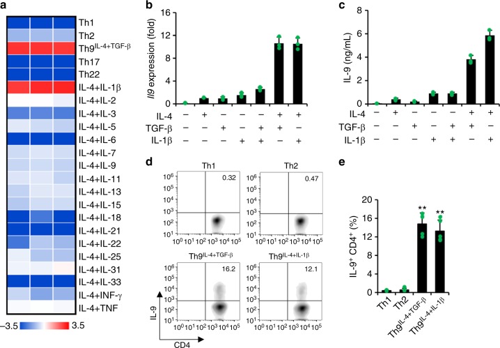
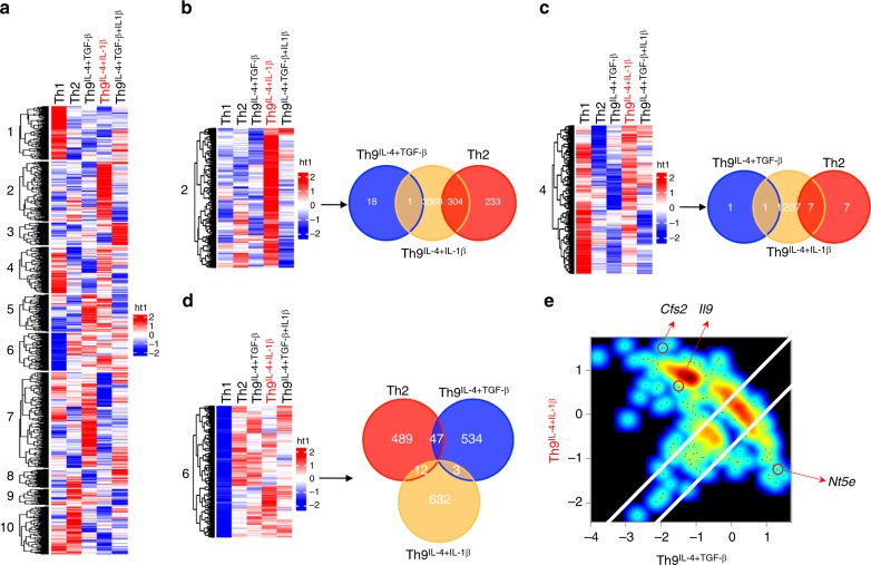
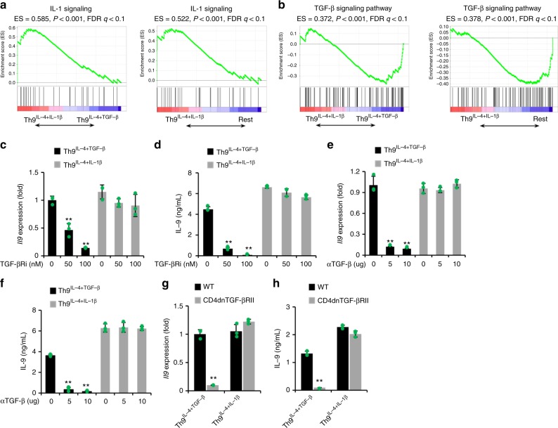
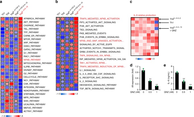
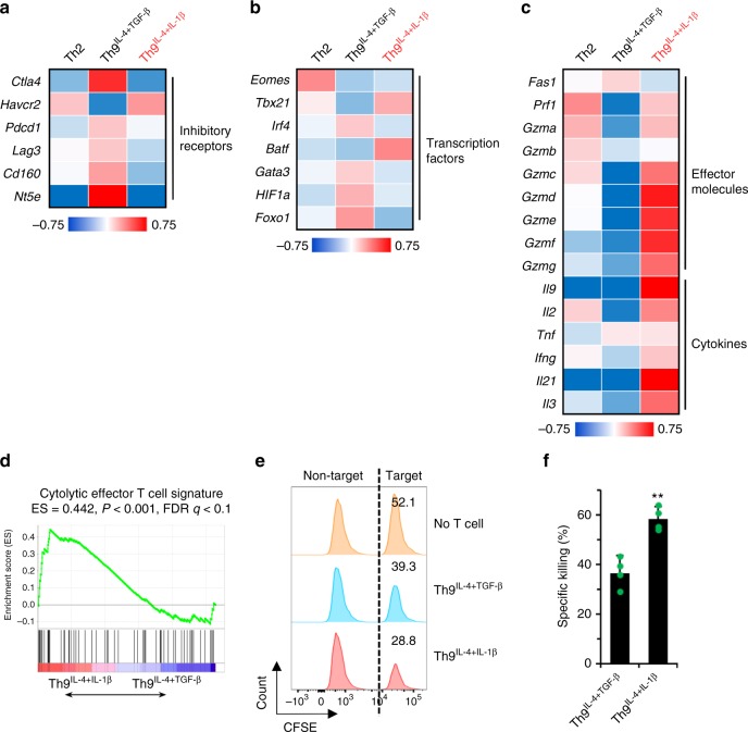
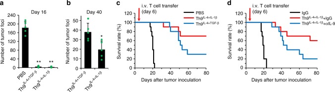
IL-9-producing CD4+ (Th9) cells are a subset of CD4+ T-helper cells that are endowed with powerful antitumor capacity. Both IL-4 and TGF-β have been reported to be indispensable for Th9 cell-priming and differentiation. Here we show, by contrast, that Th9 cell development can occur in the absence of TGF-β signaling. When TGF-β was replaced by IL-1β, the combination of IL-1β and IL-4 efficiently promoted IL-9-producing T cells (Th9IL-4+IL-1β). Th9IL-4+ IL-1β cells are phenotypically distinct T cells compared to classic Th9 cells (Th9IL-4+TGF-β) and other Th cells, and are enriched for IL-1 and NF-κB gene signatures. Inhibition of NF-κB but not TGF-β-signaling negates IL-9 production by Th9IL-4+IL-1β cells. Furthermore, when compared with classic Th9IL-4+TGF-β cells, Th9IL-4+IL-1β cells are less exhausted, exhibit cytotoxic T effector gene signature and tumor killing function, and exert a superior antitumor response in a mouse melanoma model. Our study thus describes an alternative pathway for Th9 cell differentiation and provides a potential avenue for antitumor therapies.


Conflict of interest statement

The authors declare no competing interests.

Figures

Fig. 1
Fig. 1
Interleukin (IL)-4 in combination with IL-1β promotes IL-9-producing CD4+ T cell development. Naive CD4+ CD62L+ T cells were purified from the spleens of tyrosinase-related protein-1 mice and cocultured with irradiated antigen-presenting cells under polarized conditions as detailed in the Methods section (polarized in vitro for 3 days). a Reverse transcriptase–PCR (RT-PCR) was performed to determine the expression of Il9 genes. Shown is the heatmap illustrating the relative expression of Il9 (data are log scaled) (n = 3). b RT-RCR analysis of Il9 expression in T cells primed with different cytokines as indicated (n = 3). c IL-9 production was measured by enzyme-linked immunosorbent assay in the supernatants of differentiated T cells in vitro (n = 3). d, e Intracellular staining showing the percentages of IL-9-producing cells in polarized T helper type 1 (Th1), Th2, classic Th9IL-4+ TGF-β, and Th9IL-4+IL-1β cells (n = 5). Representative data (d) and summarized results (e) are shown. Representative results of one from at least two repeated experiments are shown. Data are mean ± SD; **P < 0.01, compared with Th1 or Th2 cells, Student’s t test
Fig. 2
Fig. 2
Th9IL-4+IL-1β cells have an identifiable transcriptional signature. Naive CD4+ CD62L+ T cells were purified from the spleens of tyrosinase-related protein-1 mice and cocultured with irradiated antigen-presenting cells under polarized conditions for 3 days, and RNA was extracted for gene array analysis. a Hierarchical clustering of expression levels of 32,471 transcripts in T helper type 1 (Th1), Th2, classic Th9IL-4+TGF-β, Th9IL-4+IL-1β, and Th9IL-4+TGF-β+IL-1β cells. Cut-tree algorithm divided the transcripts into 10 clusters according to the distances between them defined by average method. b Venn diagrams displaying the number of upregulated genes of Th2, classic Th9IL-4+TGF-β, and Th9IL-4+IL-1β cells in cluster 2. The threshold for the upregulated genes is set as z-score = 0.9. c Venn diagrams showing the number of upregulated genes of Th2, classic Th9IL-4+TGF-β, and Th9IL-4+IL-1β cells in cluster 4. The threshold for the upregulated genes is set as z-score = 0.9. d Venn diagrams showing the number of upregulated genes of Th2, classic Th9IL-4+TGF-β, and Th9IL-4+IL-1β cells in cluster 6. The threshold for the upregulated genes is set as z-score = 0.9. e Specific genes upregulated or downregulated by Th9IL-4+IL-1β cells versus classic Th9IL-4+TGF-β. Axis denotes the scaled expression levels represented by z-scores in the scatter plot. Csf2 and Il9 are uniquely upregulated (z-score is >1 and 0, respectively) in Th9IL-4+IL-1β cells, whereas Nt5e is specifically upregulated in classic Th9IL-4+TGF-β cells with z-score >1
Fig. 3
Fig. 3
Differentiation of Th9IL-4+IL-1β cells is transforming growth factor (TGF)-β independent. af T cells differentiation and gene array data are the same as shown in Fig. 2. a Gene set enrichment analysis (GSEA) of Th9IL-4+IL-1β cells versus classic Th9IL-4+TGF-β cells or the Rest T helper (Th) cells for interleukin (IL)-1 signaling genes. Rest Th cells contain Th1, Th2, Th9IL-4+TGF-β, and Th9IL-4+TGF-β+IL-1β cells. b GSEA of Th9IL-4+IL-1β versus classic Th9IL-4+TGF-β cells or the Rest Th cells for TGF-β signaling genes. c, d Reverse transcriptase–PCR (RT-PCR) analysis of Il9 transcriptional level (c) and enzyme-linked immunosorbent assay (ELISA) of IL-9 production in the supernatants (d) from TGF-βRi-treated classic Th9IL-4+TGF-β cells or Th9IL-4+IL-1β cells at the indicated concentrations (n = 3). TGF-βRi TGF-β receptor serine kinase inhibitor. e, f RT-PCR analysis of Il9 transcriptional level (e) and ELISA of IL-9 production in the supernatants (f) from αTGF-β-treated classic Th9IL-4+TGF-β cells or Th9IL-4+IL-1β cells at the indicated concentrations (n = 3). αTGF-β TGF-β monoclonal antibody (mAb). g, h Naive CD4+ CD62L+ T cells were purified from the spleens of wild-type (WT) mice or CD4dnTGF-βRII mice and cultured with plate-bound anti-CD3 mAbs and soluble anti-CD28 mAbs under polarized conditions, as detailed in the Methods section, for 3 days. RT-PCR analysis of Il9 transcriptional level (g) and ELISA of IL-9 production in the supernatants (h) of classic Th9IL-4+TGF-β cells and Th9IL-4+IL-1β cells from WT mice or CD4dnTGF-βRII mice after in vitro differentiation (n = 3). CD4dnTGF-βRII mice mice expressing a dominant-negative TGF-β receptor. Data are mean ± SD; **P < 0.01, Student’s t test. Representative results of one from two repeated experiments are shown
Fig. 4
Fig. 4
Nuclear factor (NF)-κB pathway is required for Th9IL-4+IL-1β cell differentiation. T cells differentiation and gene array data are the same as shown in Fig. 2. a, b Heatmaps of enriched signaling of the most upregulated/downregulated pathways for T helper type 1 (Th1), Th2, classic Th9IL-4+TGF-β, Th9IL-4+IL-1β, and Th9IL-4+TGF-β+IL-1β cells. c Interleukin (IL)-9 production was measured by enzyme-linked immunosorbent assay (ELISA) in the supernatants of in vitro differentiated T cells. Shown is the heatmap of IL-9 relative production in the supernatants of Th1 and Th9IL-4+IL-1β cells and inhibitor-treated Th9IL-4+IL-1β cells (Kinase Inhibitor library, also see Supplementary Figure 10a). d, e RT-PCR analysis of Il9 transcriptional level (d) and ELISA of IL-9 production in the supernatants (e) of QNZ-treated Th9IL-4+IL-1β cells at the indicated concentrations (n = 3). QNZ NF-κB pathway inhibitor. Data are mean ± SD; **P < 0.01, Student’s t test. Representative results from one of two repeated experiments are shown
Fig. 5
Fig. 5
Th9IL-4+IL-1β cells exhibited antitumor activity in vitro. T cells differentiation and gene array data are the same as shown in Fig. 2. a Heatmap illustrating the relative expression of exhaustion/inhibition markers as indicated (data are log scaled). b Heatmap illustrating the relative gene expression of transcription factors as indicated (data are log scaled). c Heatmap illustrating the relative gene expression (data are log scaled). d Gene set enrichment analysis of classic Th9IL-4+TGF-β cells versus Th9IL-4+IL-1β cells for cytolytic effector T cell signature. e, f Specific killing assay of tyrosinase-related protein-1 classic Th9IL-4+TGF-β or Th9IL-4+IL-1β was performed against CFSEhigh-B16 tumor cells as target cells and CFSElow-MC38 tumor cells as a non-target control. An E:T ratio of 10:1 was used, and specific killing was determined after 48 h of coculture. Representative data (e) and summarized data (f) are shown (n = 4). Data are mean ± SD; **P < 0.01, Student’s t test. Representative results from one of two repeated experiments are shown
Fig. 6
Fig. 6
The antitumor activity of Th9IL-4+IL-1β cells in vivo. C57BL/6 mice were challenged with 1 × 105 B16 cells delivered intravenously. T cells were differentiated as in Fig. 2, and 1 × 106 tyrosinase-related protein (TRP)-1 T cells were transferred on day 6 after tumor challenge. a, b TRP-1-specific classic Th9IL-4+TGF-β cells or Th9IL-4+IL-1β cells were transferred into tumor-bearing mice, tumor foci in the lung were counted on day 16 (a) or day 40 (b) after tumor inoculation (n = 5 mice/group). c The survival rate of tumor-bearing mice with the indicated treatments (n = 10 mice). d The survival rate of tumor-bearing mice with the indicated treatments (n = 10 mice). Control IgG or αIL-9 were injected intraperitoneally twice every week starting at 1 day before intravenous transfer of T cells. Data are mean ± SD; *P < 0.05, **P < 0.01, Student’s t test. Representative results from one of two repeated experiments are shown

References

    1. Dardalhon V, et al. IL-4 inhibits TGF-β-induced Foxp3+T cells and, together with TGF-β, generates IL-9+IL-10+Foxp3- effector T cells. Nat. Immunol. 2008;9:1347–1355. doi: 10.1038/ni.1677. - DOI - PMC - PubMed
    1. Veldhoen M, et al. Transforming growth factor-β ‘reprograms’ the differentiation of T helper 2 cells and promotes an interleukin 9-producing subset. Nat. Immunol. 2008;9:1341–1346. doi: 10.1038/ni.1659. - DOI - PubMed
    1. Chang HC, et al. The transcription factor PU.1 is required for the development of IL-9-producing T cells and allergic inflammation. Nat. Immunol. 2010;11:527–534. doi: 10.1038/ni.1867. - DOI - PMC - PubMed
    1. Staudt V, et al. Interferon-regulatory factor 4 is essential for the developmental program of T helper 9 cells. Immunity. 2010;33:192–202. doi: 10.1016/j.immuni.2010.07.014. - DOI - PubMed
    1. Goswami R, et al. STAT6-dependent regulation of Th9 development. J. Immunol. 2012;188:968–975. doi: 10.4049/jimmunol.1102840. - DOI - PMC - PubMed

Publication types

MeSH terms