. 2019 Mar 26;9(1):5168.
doi: 10.1038/s41598-019-41490-w.

Developing a blood-based gene mutation assay as a novel biomarker for oesophageal adenocarcinoma

Affiliations

Developing a blood-based gene mutation assay as a novel biomarker for oesophageal adenocarcinoma

Hasan N Haboubi et al. Sci Rep. .

Abstract

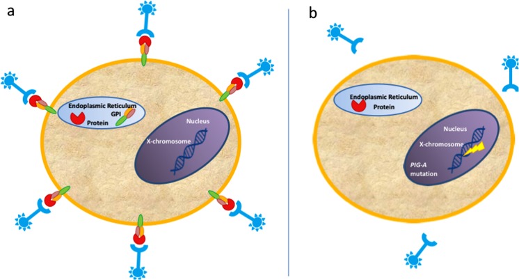
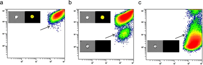
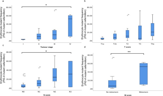
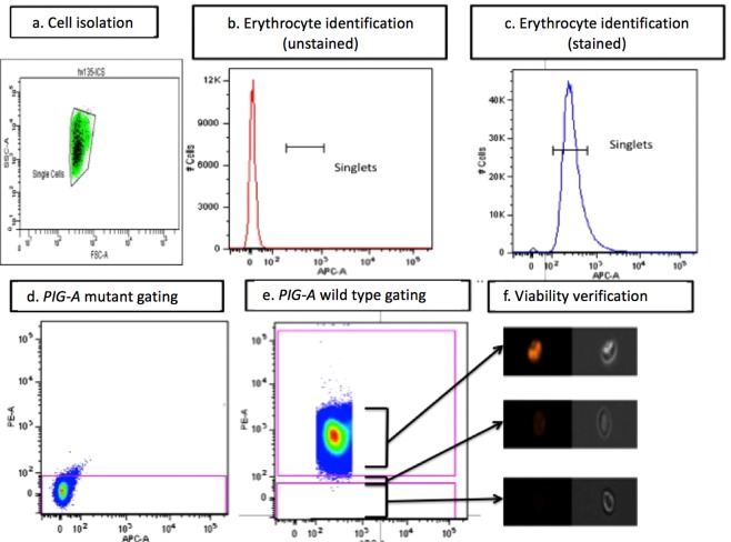
The Phosphatidylinositol glycan class A (PIG-A) gene mutation assay phenotypically measures erythrocyte mutations, assessed here for their correlation to neoplastic progression in the gastro-oesophageal reflux disease (GORD)-Barrett's metaplasia (BM)-oesophageal adenocarcinoma (OAC) model. Endoscopy patients underwent venipuncture and erythrocytes fluorescently stained for glycosyl phosphatidylinositol (GPI)-anchored proteins; CD55 and CD59. Using flow cytometry, GPI-anchor negative erythrocytes (mutants) were scored and compared amongst groups. The study enlisted 200 patients and 137 healthy volunteers. OAC patients had a three-fold increase in erythrocyte mutant frequency (EMF) compared to GORD patients (p < 0.001) and healthy volunteers (p < 0.001). In OAC patients, higher EMF was associated with worsening tumour staging (p = 0.014), nodal involvement (p = 0.019) and metastatic disease (p = 0.008). Chemotherapy patients demonstrated EMF's over 19-times higher than GORD patients. Patients were further classified into groups containing those with non-neoplastic disease and those with high-grade dysplasia/cancer with 72.1% of cases correctly classified by high EMF. Within the non-neoplastic group, aspirin users had lower EMF (p = 0.001) and there was a positive correlation between body mass index (p = 0.03) and age (p < 0.001) and EMF. Smokers had EMF's over double that of non-smokers (p = 0.011). Results suggest this test could help detect OAC and may be a useful predictor of disease progression.


Conflict of interest statement

The authors declare no competing interests.

Figures

Figure 1
Figure 1
The phosphatidylinositol glycan class A (PIG-A) gene codes for a critical step in the formation of glycosyl phosphatidylinositol (GPI)-Anchors. (a) Intact PIG-A gene allows for GPI-anchor synthesis and presentation of proteins on cell surface membrane, detectable using fluorescently tagged antibodies and flow cytometric methodologies. (b) Silencing Mutations to the PIG-A gene lead to absence of GPI-anchored proteins and non-fluorescent phenotypic mutants.
Figure 2
Figure 2
Mutant and wild-type phosphatidylinositol glycan class A (Pig-A) L5178Y cells. Mutant cells negative for the glycosyl phosphatidylinositol (GPI)-linked protein CD90.2 are indicated by arrows. (a) Untreated L5178Y cells. (b) L5178Y’s treated with 2 mM ethyl methanesulphonate (EMS) and enriched using anti-PE magnetic beads. (c) Mutagenized L5178Y cells sorted using fluorescent activated cell sorting. Insert: Image stream analysis of cells displaying brightfield (left) and CD90.2-PE fluorescent image (right) for Pig-A wild-type (top) and mutant cell (bottom).
Figure 3
Figure 3
Erythrocyte mutation frequencies measured in different histological subsets; healthy volunteers (n = 137) together with patients with gastro-oesophageal reflux disease (GORD) (n = 77), non-dysplastic Barrett’s metaplasia (NDBM) (n = 62), low-grade dysplasia (LGD) (n = 4), high-grade dysplasia (HGD) (n = 7) and oesophageal adenocarcinoma who had not undergone chemotherapy (OAC) (n = 42). A significant increase in EMF in OAC patients compared to those with NDBM, GORD and healthy volunteers (HV)(*p < 0.05, **p < 0.01, ***p < 0.001).
Figure 4
Figure 4
Erythrocyte mutant frequency (EMF) scores for different tumour staging further broken down into aspects of Tumour Node Metastasis (TNM) scoring. Statistically determined outliers are represented with their corresponding participant ID as either a circle or asterix. The number of patients in each staging subgroup is provided below. (a) Significant increase in EMF with increasing tumour staging (p = 0.014). TI (n = 5), TII (n = 19), TIII (n = 12) and TIV (n = 6). (b) No difference in EMF between different primary tumour sizes (T-score) (p = 0.627). T1a (n = 3), T1b (n = 8), T2 (n = 16), T3 (n = 12) and T4a (n = 3). (c) A significant increase in EMF with more lymph node involvement (N-score). (p = 0.019). N0 (n = 15), N1 (n = 14), N2 (n = 11) and N3 (n = 2). (d) Higher EMF significantly associated with metastatic disease (M-score)(p = 0.008). M0 (n = 36) and M1 (n = 6).
Figure 5
Figure 5
Gating Strategies employed for erythrocyte mutant frequency (EMF) analysis. (a) Erythrocytes initially identified by their forward scatter and side scatter profiles. (b) CD235a staining is used to characterize red blood cells and exclude other cell populations. (c) Single cells can be identified separate to clumped cells and gated for subsequent analysis. (d) A gate is drawn from an instrument calibration standard (ICS) to enable isolation of mutant (negatively fluorescent) cells. (e) Analysis of double stained samples reveal positively stained wild type red blood cells, and negative mutant populations. (f) Gating can be performed using cytograms and images viewed using the imagestream (Amnis) to confirm fluorescent status and cell morphology.

References

    1. Lagergren J, Bergström R, Lindgren A, Nyrén O. Symptomatic Gastroesophageal Reflux as a Risk Factor for Esophageal Adenocarcinoma. N. Engl. J. Med. 1999;340:825–831. doi: 10.1056/NEJM199903183401101. - DOI - PubMed
    1. Spechler SJ, Zeroogian JM, Antonioli DA, Wang HH, Goyal RK. Prevalence of metaplasia at the gastro-oesophageal junction. Lancet. 1994;344:1533–1536. doi: 10.1016/S0140-6736(94)90349-2. - DOI - PubMed
    1. Desai TK, et al. The incidence of oesophageal adenocarcinoma in non-dysplastic Barrett’s oesophagus: a meta-analysis. Gut. 2012;7:970–976. doi: 10.1136/gutjnl-2011-300730. - DOI - PubMed
    1. Dubecz A, et al. Does the Incidence of Adenocarcinoma of the Esophagus and Gastric Cardia Continue to Rise in the Twenty-First Century?-a SEER Database Analysis. J. Gastrointest. Surg. 2014;18:124–129. doi: 10.1007/s11605-013-2345-8. - DOI - PubMed
    1. Zhang Y. Epidemiology of esophageal cancer. World Journal of Gastroenterology. 2013;19:5598–5606. doi: 10.3748/wjg.v19.i34.5598. - DOI - PMC - PubMed

Publication types

MeSH terms

Substances

Supplementary concepts