Review
. 2019 Mar 11:9:1401.
doi: 10.3389/fphar.2018.01401. eCollection 2018.

Carbon-Based Nanomaterials for Biomedical Applications: A Recent Study

Affiliations
Review

Carbon-Based Nanomaterials for Biomedical Applications: A Recent Study

Debabrata Maiti et al. Front Pharmacol. .

Abstract

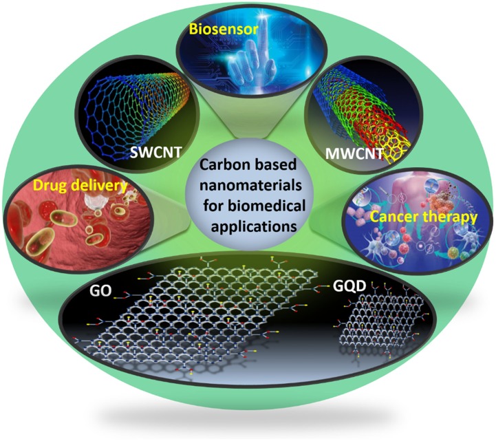
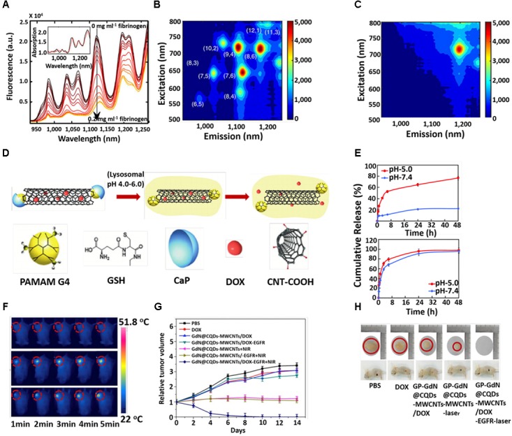
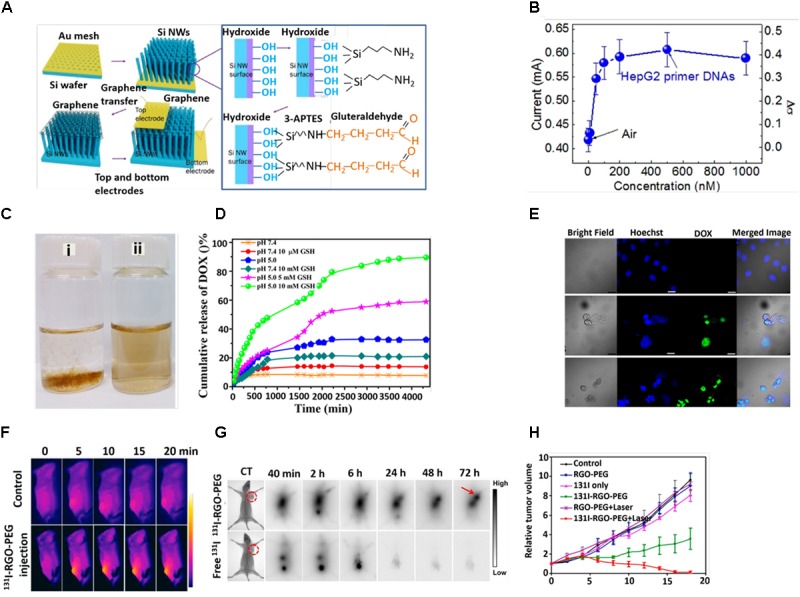
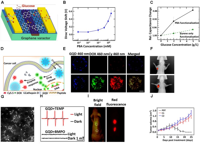
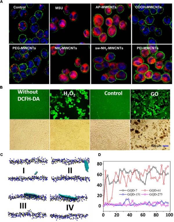
The study of carbon-based nanomaterials (CBNs) for biomedical applications has attracted great attention due to their unique chemical and physical properties including thermal, mechanical, electrical, optical and structural diversity. With the help of these intrinsic properties, CBNs, including carbon nanotubes (CNT), graphene oxide (GO), and graphene quantum dots (GQDs), have been extensively investigated in biomedical applications. This review summarizes the most recent studies in developing of CBNs for various biomedical applications including bio-sensing, drug delivery and cancer therapy.

Keywords: biomedical applications; biosensor; cancer therapy; carbon nanomaterials; drug delivery.


Figures

FIGURE 1
FIGURE 1
Schematic illustration for the biomedical applications of carbon-based nanomaterials (CBNs).
FIGURE 2
FIGURE 2
(A) Fluorescence spectra of 1,2-bis(diphenylphosphino) ethane (DPPE)-polyethylene glycol (PEG)-SWCNT with different concentrations of fibrinogen. Excitation-emission of DPPE-PEG-SWCNT solution (B) before and after (C) fibrinogen adding. (D) Schematic illustration for the triggered release of DOX from DOX-loaded CaP nanocapsule under intracellular endo/lysosomal conditions. (E) DOX release profile at pH 7.4 and 5 with time from CNT–G4–GSH–CaP–DOX and (upper) CNT–G4–GSH–DOX (lower). (F) In vivo photothermal images under 5 min NIR laser (808 nm, 1 W/cm2) irradiation. (G) Tumor growth curves after different treatments at different times. (H) Digital photographs of tumors and tumor-bearing mice after different treatments. Copyright Bisker et al. (2016) Nature publishing group, Banerjee et al. (2015) Royal Society of Chemistry, and Zhang et al. (2017) Elsevier.
FIGURE 3
FIGURE 3
(A) Schematic diagram of fabrication process. (B) Responsivity of graphene/Si-NWs biosensors: Current of a graphene/Si-NWs biosensor as a function of mole fraction of p-ss oligonucleotide. (C) Digital photographs of aqueous dispersion of (i) GON and (ii) GON-Cy-ALG-PEG in PBS. (D) DOX release profile at pH 7.4 and 5 in presence and absence of GSH. (E) Cellular uptake of HepG2 cells stained by Hoechst (blue), DOX (green)-loaded GON-Cy-ALG-PEG in presence and absence of GSH. Bars represent 30 μm. (F) In vivo photothermal therapy (G) in vivo gamma imaging study. (H) Tumor volume of mice after treatment. Copyright Kim et al. (2016) Nature publishing group, Zhao et al. (2015) American Chemical Society, and Chen et al. (2015) Elsevier.
FIGURE 4
FIGURE 4
(A) Schematic diagram of GQD-based varactor for glucose sensing. (B) Dirac voltage shift of graphene varactors with different concentrations of PBA solutions. (C) Curves for change in relative capacitance with glucose concentrations. (D) Schematic diagram for the drug delivery and release of GQD-based theranostic agent. (E) CLSM images of the multicellular tumor spheroids incubation with GQD-P-Cy and DOX@GQD-P-Cy. Scale bar: 100 μm. (F) In vivo fluorescence images of 4T1 tumor bearing mice after intravenous treatment with DOX@GQD-P-Cy. (G) STEM images. Scale bar, 20 nm. (H) The electron spin resonance (ESR) signals of 1O2 (up) and reactive oxygen species (ROS) (down) generated upon irradiation of GQDs for 8 min in the presence of 2,2,6,6-tetramethylpiperidine and 5-tert-butoxycarbonyl-5-methyl-1-pyrroline N-oxide, respectively. (I) In vivo fluorescence images of GQDs. (J) Time dependent tumor growth curves after different treatments. Copyright Ding et al. (2017) and Zhang D.Y. et al. (2017); American Chemical Society and Ge et al. (2014) Nature publishing group.
FIGURE 5
FIGURE 5
(A) Visualization of cathepsin B localization in THP-1 cells exposed to tubes. Lysosomal damage and cathepsin B release were identified by using Magic Red staining. THP-1 cells were seeded into 8-well chamber slides and incubated with f-MWCNTs at 120 μg/ml in complete RPMI 1640 for 3 h. After fixation, cells were stained with Magic Red (ImmunoChemistry Technologies), wheat germ agglutinin-Alexa 488, and Hoechst 33342 dye, followed by visualization under a confocal 1P/FCS inverted microscope. (B) ROS production in GLC-82 cells treated with 100 mg/L of GO for 48 h. The positive control was prepared by culturing the cells with RPMI-1640 containing 100 μM of H2O2 for 1 h prior to the addition of DCFH-DA. The cells without DCFH-DA treatment was taken as a negative control. The control means that cells without exposure to GO were labeled by the DCFH-DA. (C) GQDs with different sizes on the membrane after 100 ns MD simulation: (I) GQD7-small size, (II) GQD61-small size, (III) GQD151-large size, and (IV) GQD275-large size. The GQDs are shown by a VDW model with VMD. N atoms (blue) and P atoms (yellow) in the membrane are also shown in the VDW model. (D) The angles between different GQDs and the x–y plane of the lipid membrane as a function of simulation time. Copyright Li et al. (2013, and Liang et al. (2016) American Chemical Society.

References

    1. Adeli M., Soleyman R., Beiranvanda Z., Madani F. (2013). Carbon nanotubes in cancer therapy: a more precise look at the role of carbon nanotube-polymer interactions. Chem. Soc. Rev. 42 5231–5256. 10.1039/c3cs35431h - DOI - PubMed
    1. Akhavan O., Ghaderi E., Rahighi R. (2012). Toward single-DNA electrochemical biosensing by graphene nanowalls. ACS Nano 6 2904–2916. 10.1021/nn300261t - DOI - PubMed
    1. Aldieri E., Fenoglio I., Cesano F., Gazzano E., Gulino G., Scarano D. (2013). The role of iron impurities in the toxic effects exerted by short multiwalled carbon nanotubes (MWCNT) in murine alveolar macrophages. J. Toxicol. Environ. Health A 76 1056–1071. 10.1080/15287394.2013.834855 - DOI - PubMed
    1. Allegri M., Perivoliotis D. K., Bianchi M. G., Chiu M., Pagliaro A., Koklioti M. A. (2016). Toxicity determinants of multi-walled carbon nanotubes: the relationship between functionalization and agglomeration. Toxicol. Rep. 3 230–243. 10.1016/j.toxrep.2016.01.011 - DOI - PMC - PubMed
    1. Alshehri R., Ilyas A. M., Hasan A., Arnaout A., Ahmed F., Memic A. (2016). Carbon nanotubes in biomedical applications: factors, mechanisms, and remedies of toxicity. J. Med. Chem. 59 8149–8167. 10.1021/acs.jmedchem.5b01770 - DOI - PubMed

LinkOut - more resources