. 2019 Mar 12:10:205.
doi: 10.3389/fneur.2019.00205. eCollection 2019.

Neuronal ICAM-5 Plays a Neuroprotective Role in Progressive Neurodegeneration

Affiliations

Neuronal ICAM-5 Plays a Neuroprotective Role in Progressive Neurodegeneration

Katharina Birkner et al. Front Neurol. .

Abstract

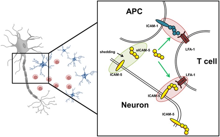
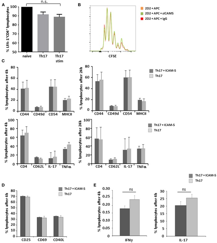
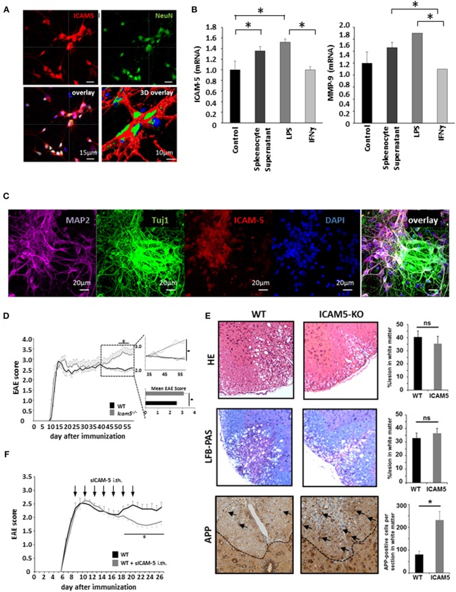
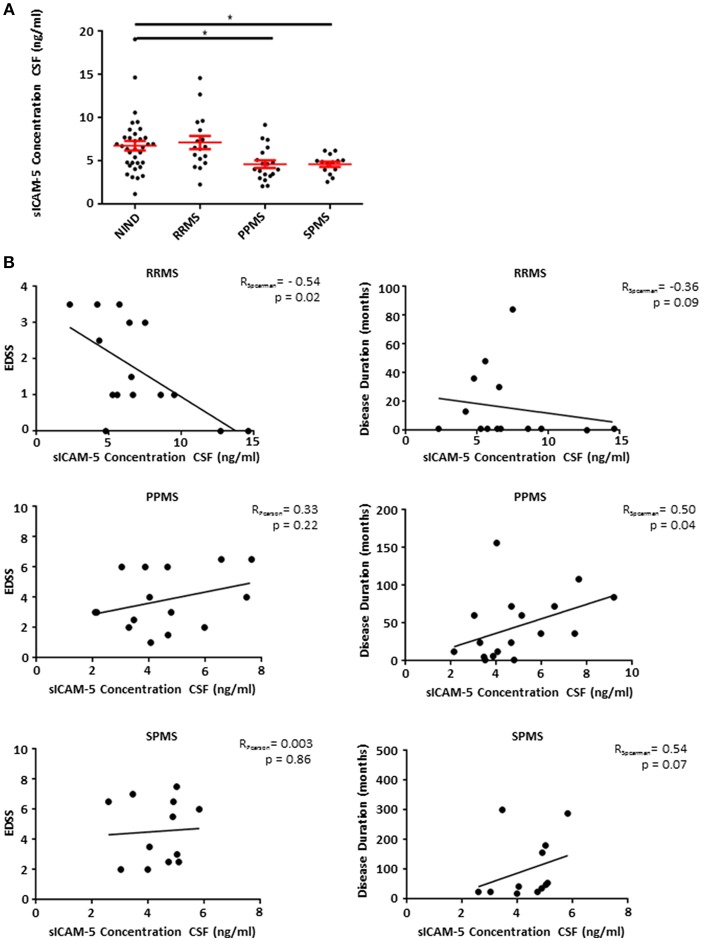
Multiple sclerosis (MS) is a chronic autoimmune disease of the central nervous system (CNS) leading to CNS inflammation and neurodegeneration. Current anti-inflammatory drugs have only limited efficacy on progressive neurodegenerative processes underlining the need to understand immune-mediated neuronal injury. Cell adhesion molecules play an important role for immune cell migration over the blood-brain barrier whereas their role in mediating potentially harmful contacts between invading immune cells and neurons is incompletely understood. Here, we assess the role of the CNS-specific neuronal adhesion molecule ICAM-5 using experimental autoimmune encephalomyelitis (EAE), an animal model of MS. ICAM-5 knockout mice show a more severe EAE disease course in the chronic phase indicating a neuroprotective function of ICAM-5 in progressive neurodegeneration. In agreement with the predominant CNS-specific function of ICAM-5, lymphocyte function-associated antigen 1 (LFA-1)/ICAM-1 contact between antigen-presenting cells and T helper (Th)17 cells in EAE is not affected by ICAM-5. Strikingly, intrathecal application of the shed soluble form, sICAM-5, ameliorates EAE disease symptoms and thus might serve locally as an endogenous neuronal defense mechanism which is activated upon neuroinflammation in the CNS. In humans, cerebrospinal fluid from patients suffering from progressive forms of MS shows decreased sICAM-5 levels, suggesting a lack of this endogenous protective pathway in these patient groups. Overall, our study points toward a novel role of ICAM-5 in CNS autoinflammation in progressive EAE/MS.

Keywords: T cells; adhesion molecules; experimental autoimmune encephalomyelitis; multiple sclerosis; neuroinflammation.


Figures

Figure 1
Figure 1
Schematic overview of potential ICAM-5 dependent T cell—neuron and T cell—APC interactions in the CNS. Schematic illustration of the interplay of neurons, T cells, and APCs in the CNS. The adhesion molecule ICAM-5 (yellow) is exclusively expressed by neurons and can be cleaved by MMP-2 and 9 from the neuronal surface (also called shedding). The soluble form (sICAM-5) has been proposed to act as an inhibitor of ICAM1–LFA-1 interactions between T cells and APCs and T cells and neurons therefore displaying a protective function. Green highlights and arrows represent anti-inflammatory/ protective function and red highlights represent proinflammatory properties.
Figure 2
Figure 2
sICAM-5 does not affect Th17—APC interactions. (A) FACS staining for LFA-1 of naïve T-cells, Th17 cells, and Th17 cells after 24 h of stimulation with anti-CD3/CD28 (n = 3 for each condition). (B) Representative depiction of CFSE stainings of naïve 2d2 T cells cocultured with APCs and sICAM-5 (10 μg/ml) or IgG (10 μg/ml) for 72 h. (C) FACS staining of Th17 cells stimulated for 4 and 24 h with anti-CD3/CD28 and with or without ICAM-5 treatment (10 μg/ml). (D,E) Naïve T cells were differentiated into Th17 cells in the presence of or without sICAM-5 treatment (10 μg/ml) and stained for extracellular (D) and intracellular (E) markers. Data shown are mean ± SEM. (A) One-way ANOVA followed by Tukey's multiple comparison test, n.s. no significance; (C–E) Student's t-test.
Figure 3
Figure 3
Absence of ICAM-5 worsens disease progression which can be reversed by application of sICAM-5. (A) Immunohistochemical staining of ICAM-5-AF647 on cortical neurons (scale bar = 15 μm, 3D overlay scale bar = 10 μm). Co-staining was performed with NeuN-AF488 and DAPI (blue). (B) mRNA analysis of ICAM-5 and MMP-9 were performed with cortical neurons after stimulation with LPS, IFNγ, and splenocyte supernatant (n = 6 for each condition). Unstimulated cortical neurons served as a control. (C) Immunohistochemical staining of MAP2- AF647, Tuj1-FITC, ICAM-5-AF568, and DAPI on cortical neurons (scale bar = 20 μm. (D) EAE was induced actively in ICAM-5-KO mice and WT littermates via the injection of MOG35−55 (two independent EAEs, untreated; WT n = 12, KO n = 12). The dotted box on the plot provides a closer look at the chronic phase of the EAE but does not agree with the x axis of the upper plot. The lower plot shows only the mean disease score of the period from day 40 after immunization. (E) EAE lesions were stained from wt (n = 9) and ICAM5 KO mice (n = 6) for inflammation (HE staining), demyelination (LFB-PAS), and axonal loss (APP) and were quantified accordingly. (F) EAE was induced actively in B6 mice via the injection of MOG35−55 and mice were treated with Methylprednisolone for 5 days as soon as the mice reached a clinical score of 2. Additionally, once animals reached a clinical score of 2, 0.2 μg ICAM-5 D1-2 Fc or human IgG was applied via intrathecal (i.th) injection by lumbar puncture seven times on every second day (WT n = 7, KO n = 7). Data shown are mean ± SEM. (C, D) Mann-Whitney U-test; *p < 0.05.
Figure 4
Figure 4
Patients suffering from progressive forms of MS show lower levels of sICAM-5 in CSF. (A) ICAM-5 concentration in ng/ml in the cerebrospinal fluid of NIND patients (NIND, n = 35), RRMS (n = 17), PPMS (n = 19), and SPMS (n = 12) patients. Data shown are mean ± SEM. One-way ANOVA followed by Tukey's multiple comparison test, *p < 0.05. (B) Correlation between EDSS or disease duration in months and ICAM-5 concentration in CSF (ng/ml). Data was tested for normality using Shapiro-Wilk normality test and Pearson's or Spearman's r and p-values were determined accordingly.
Figure 5
Figure 5
No significant correlation between neurofilament light chain levels and sICAM-5. (A) ICAM-5 was correlated with NfL concentrations (pg/ml) in the cerebrospinal fluid and the serum of SPMS patients (n = 7–8). (B) ICAM-5 was correlated to NfL concentrations (pg/ml) in the cerebrospinal fluid and the serum of PPMS patients (n = 7–8). (C) ICAM-5 was correlated to NfL concentrations (pg/ml) in the cerebrospinal fluid and the serum of pooled SPMS and PPMS patients (n = 14–16). For all figures: Data was tested for normality using Shapiro-Wilk normality test and Pearson's or Spearman's r and p-values was determined accordingly.

References

    1. Cua DJ, Sherlock J, Chen Y, Murphy CA, Joyce B, Seymour B, et al. Interleukin-23 rather than interleukin-12 is the critical cytokine for autoimmune inflammation of the brain. Nature. (2003) 421:744–8. 10.1038/nature01355 - DOI - PubMed
    1. Langrish CL, Chen Y, Blumenschein WM, Mattson J, Basham B, Sedgwick JD, et al. IL-23 drives a pathogenic T cell population that induces autoimmune inflammation. J Exp Med. (2005) 201:233–40. 10.1084/jem.20041257 - DOI - PMC - PubMed
    1. Komiyama Y, Nakae S, Matsuki T, Nambu A, Ishigame H, Kakuta S, et al. IL-17 plays an important role in the development of experimental autoimmune encephalomyelitis. J Immunol. (2006) 177:566–73. - PubMed
    1. Bittner S, Wiendl H. Neuroimmunotherapies Targeting T Cells: from pathophysiology to therapeutic applications. Neurotherapeutics. (2016) 13:4–19. 10.1007/s13311-015-0405-3 - DOI - PMC - PubMed
    1. Ellwardt E, Walsh JT, Kipnis J, Zipp F. Understanding the Role of T Cells in CNS Homeostasis. Trends Immunol. (2016) 37:154–65. 10.1016/j.it.2015.12.008 - DOI - PubMed

LinkOut - more resources