. 2019 Mar 11:10:418.
doi: 10.3389/fmicb.2019.00418. eCollection 2019.

Rapid and Specific Detection of All Known Nipah virus Strains' Sequences With Reverse Transcription-Loop-Mediated Isothermal Amplification

Affiliations

Rapid and Specific Detection of All Known Nipah virus Strains' Sequences With Reverse Transcription-Loop-Mediated Isothermal Amplification

Liping Ma et al. Front Microbiol. .

Abstract

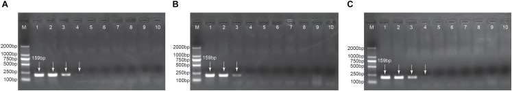
Nipah virus (NiV) is a zoonotic virus and can be transmitted through contaminated food or directly between people. NiV is classified as a Biosafety Level 4 agent, not only because of its relatively high case fatality rate, but also because there is no vaccine or other medical countermeasures and it appears to be transmitted by fomites/particulates. The development of rapid detection assay for NiV is of great importance because no effective field test is currently available. In this study, an isothermal (65°C) reverse transcription-loop-mediated isothermal amplification (RT-LAMP) method was developed, targeting the nucleocapsid protein (N) gene, for the rapid detection of NiV, and was compared with conventional RT-PCR. Three pseudoviruses of NiV N gene representing all known strains were constructed to replace live NiV. A set of RT-LAMP primers, targeting a highly conserved region of the N gene in the viral genome was designed to identify all known NiV strains. Sensitivity tests indicated that the detection limit of the RT-LAMP assay was approximately 100 pg of total NiV pseudovirus RNA, which is at least 10-fold higher than that of conventional RT-PCR. Specificity tests showed that there was no cross-reactivity with nucleocapsid protein gene of Hendra virus, Newcastle disease virus, Japanese encephalitis virus, or Influenza A virus. The RT-LAMP assay provides results within 45 min, and requires no sophisticated instruments, except an isothermal water bath or metal bath with 1 μl calcein indicator. An analysis of the clinical samples showed that the assay had good stability. In conclusion, systematic experiments have shown that the RT-LAMP assay developed here effectively detects three NiV pseudoviruses representing all known strains of NiV, with high specificity, sensitivity and stability.

Keywords: N gene; Nipah virus; RT-LAMP; rapid detection; reverse transcription-loop-mediated isothermal amplification.


Figures

FIGURE 1
FIGURE 1
Sequence alignment of the conserved region of NiV N gene targeted for RT-LAMP detection. The target regions of each representative genotype strain were aligned. Nucleotides identical to those of NiV Bangladeshi isolate (GenBank accession JN808862.1) are replaced with dots. The primer sequences are shown below the aligned sequence, and the positions are numbered based on the complete N gene of Bangladeshi isolate (GenBank accession JN808862.1). Outer primers, inner primers (BIP and FIP primers are defined), and loop primers are shown with green arrows, blue arrows, and red arrows, respectively. The colors of the four bases differ, and the nine degenerate bases are not colored.
FIGURE 2
FIGURE 2
Selection of optimal reaction conditions. Panel A shows the turbidity signals of the three candidate primer sets, which indicate that the set two primers were optimal. This set of primers was used in all later studies. Panel B shows the temperature selection results, and the turbidity at each temperature is shown in a different color.
FIGURE 3
FIGURE 3
The specificity of the RT-LAMP assay. Panel A shows the turbidity results of the specificity test of NiV. The positive controls were the three NiV N gene plasmids pUC57-NiV-BN (Bangladeshi isolate), pUC57-NiV-MN (Malaysian isolate), and pUC57-NiV-TN (N gene sequence from Thai bat RNAs), marked as LAMP-BN, LAMP MN, and LAMP-TN, respectively. LAMP detection of pET-HeV is marked as LAMP-HeV. RT-LAMP detections of the three pseudoviruses (pha-NiV-BN, pha-NiV-MN, and pha-NiV-TN) and the RNA of pET-HeV, Newcastle Disease virus (NDV), Japanese encephalitis virus vaccine strain SA14-14-2 (JEV), and Influenza A virus A/Puerto Rico/8(H1N1) (PR8) are labeled RT-LAMP-BN, RT-LAMP-MN, RT-LAMP-TN, RT-LAMP-HeV, RT-LAMP-NDV, RT-LAMP-JEV, and RT-LAMP-PR8, respectively. Negative controls were the RNA from the cell extract transfected with pHAGE-CMV-MCS-IZsGreen vector and nuclease-free water, and are marked as RT-NC-CMV and RT-NC-NF, respectively. Panel B shows the specificity of the assay, using detection with 1 μl of calcein indicator. Numbers 1–13 represent LAMP-BN, LAMP-MN, LAMP-TN, RT-LAMP-BN, RT-LAMP-MN, RT-LAMP-TN, LAMP-HeV, RT-LAMP-HeV, RT-LAMP-NDV, RT-LAMP-JEV, RT-LAMP-PR8, RT-NC-CMV, and RT-NC-NF, respectively. Positive samples are marked “+” and negative samples are marked “−.”
FIGURE 4
FIGURE 4
Sensitivity of the RT-LAMP assay. Panels A–C show the turbidity in the RT-LAMP sensitivity tests for pha-NiV-BN, pha-NiV-MN, and pha-NiV-TN, respectively. LAMP-50 ng/μl of each panel represents the positive controls: pUC57-NiV-BN (50 ng/μl), pUC57-NiV-MN (50 ng/μl), and pUC57-NiV-TN (50 ng/μl), respectively. Negative control (RNA from the cell extract transfected with pHAGE-CMV-MCS-IZsGreen vector) in each panel is marked with NC-CMV and the concentrations tested in each RT-LAMP reaction are listed on the right of each panel. Panels D–F show the chromogenic test results for the RT-LAMP detection of pha-NiV-BN, pha-NiV-MN, and pha-NiV-TN, respectively, with 1 μl of calcein indicator. Numbers on the tubes of each panel correspond to the legends in panels A–C, respectively. Positive results are marked “+” and negative results are marked “−.”
FIGURE 5
FIGURE 5
Detection limits of conventional RT-PCR. Panels A–C show the RT-PCR detection limits of pha-NiV-BN, pha-NiV-MN, and pha-NiV-TN, respectively. Lane M: DL2000 DNA Marker was used as the size marker; lane 1: positive controls, pUC57-NiV-BN, pUC57-NiV-MN, and pUC57-NiV-TN in panels A–C, respectively; lanes 2–6: 10−1–10−6 dilutions of the cDNA of pha-NiV-BN, pha-NiV-MN, and pha-NiV-TN, respectively; lanes 8–9 in the three panels: cDNA from the cell extract transfected with pHAGE-CMV-MCS-IZsGreen vector and nuclease-free water, respectively (negative controls). The lengths of products are labeled in lane 1 of each panel. Positive results are indicated with white arrows.
FIGURE 6
FIGURE 6
Stability of the RT-LAMP assay in clinical samples. Panel A–C show the turbidity-based RT-LAMP detection limits for pha-NiV-BN, pha-NiV-MN, and pha-NiV-TN, respectively, in clinical samples. The four diagrams, from left to right in each panel, represent the blood clinical sample, fecal clinical sample, throat swab clinical sample, and urine sample, which are abbreviated “B,” “F,” “TS,” and “U,” respectively. “n” and “p” in panels A–C represent “ng/μl” and “pg/μl,” respectively. The negative controls (RNA from the cell extract transfected with pHAGE-CMV-MCS-IZsGreen vector and the genomic solutions extracted from four common clinical samples taken of the same normal person) in each panel are marked NC-CMV, NC-B, NC-F, NC-TS, and NC-U, respectively.

References

    1. Arankalle V. A., Bandyopadhyay B. T., Ramdasi A. Y., Jadi R., Patil D. R., Rahman M., et al. (2011). Genomic characterization of Nipah virus, West Bengal, India. Emerg. Infect. Dis. 17 907–909. 10.3201/eid1705.100968 - DOI - PMC - PubMed
    1. Cao Z., Wang H., Wang L., Li L., Jin H., Xu C., et al. (2016). Visual detection of west nile virus using reverse transcription loop-mediated isothermal amplification combined with a vertical flow visualization strip. Front. Microbiol. 7:554. 10.3389/fmicb.2016.00554 - DOI - PMC - PubMed
    1. Chadha M. S., Comer J. A., Lowe L., Rota P. A., Rollin P. E., Bellini W. J., et al. (2006). Nipah virus–associated encephalitis outbreak, Siliguri, India. Emerg. Infect. Dis. 12 235–240. 10.3201/eid1202.051247 - DOI - PMC - PubMed
    1. Chadha M. S., Comer J. A., Lowe L., Rota P. A., Rollin P. E., Bellini W. J., et al. (2016). Nipah virus-associated encephalitis outbreak, Siliguri, India. Emerg. Infect. Dis. 12 235–240. 10.3201/eid1202.051247 - DOI - PMC - PubMed
    1. Chow V. T., Tambyah P. A., Yeo W. M., Phoon M. C., Howe J. (2000). Diagnosis of Nipah virus encephalitis by electron microscopy of cerebrospinal fluid. J. Clin. Virol. 19 143–147. 10.1016/S1386-6532(00)00094-9 - DOI - PubMed

LinkOut - more resources