. 2019 May 21;47(9):4707-4720.
doi: 10.1093/nar/gkz208.

The molecular mechanism of dsRNA processing by a bacterial Dicer

Affiliations

The molecular mechanism of dsRNA processing by a bacterial Dicer

Lan Jin et al. Nucleic Acids Res. .

Abstract

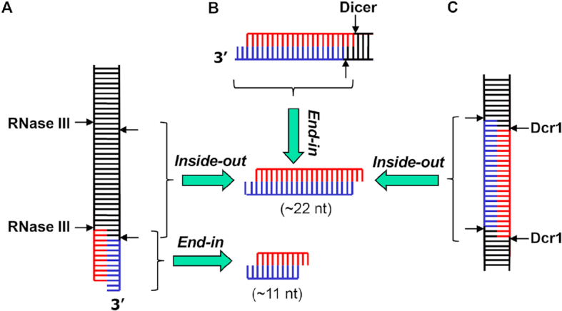
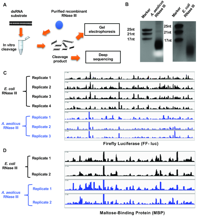
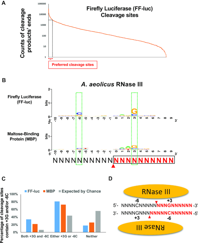
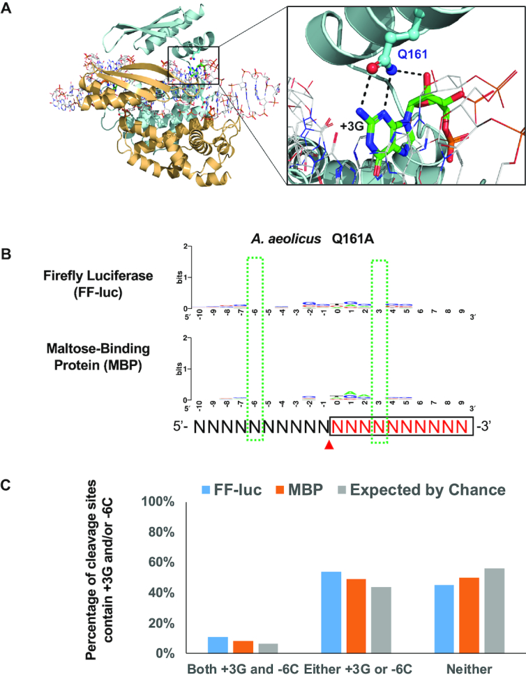
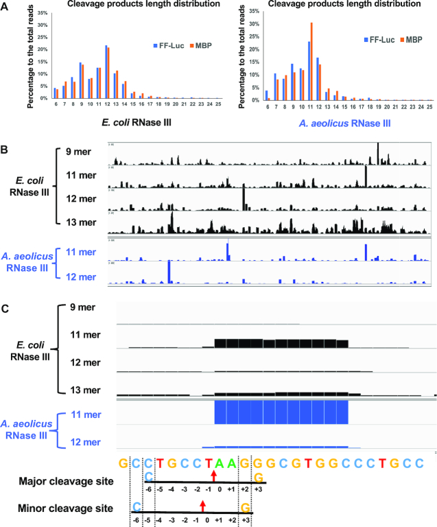
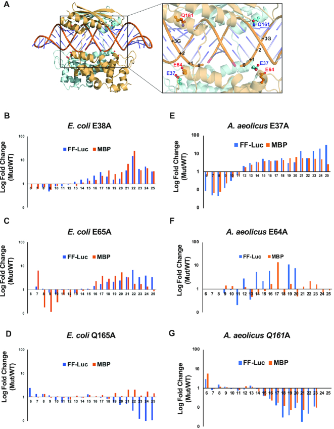
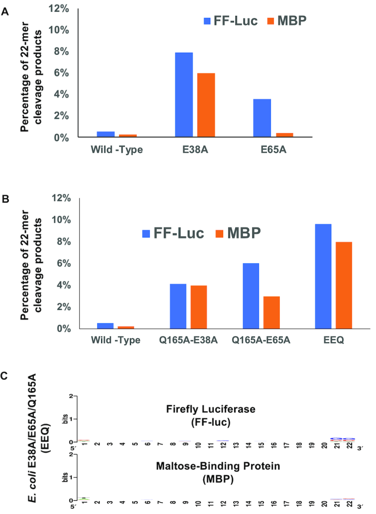
Members of the ribonuclease (RNase) III family regulate gene expression by processing dsRNAs. It was previously shown that Escherichia coli (Ec) RNase III recognizes dsRNA with little sequence specificity and the cleavage products are mainly 11 nucleotides (nt) long. It was also shown that the mutation of a glutamate (EcE38) to an alanine promotes generation of siRNA-like products typically 22 nt long. To fully characterize substrate specificity and product size of RNase IIIs, we performed in vitro cleavage of dsRNAs by Ec and Aquifex aeolicus (Aa) enzymes and delineated their products by next-generation sequencing. Surprisingly, we found that both enzymes cleave dsRNA at preferred sites, among which a guanine nucleotide was enriched at a specific position (+3G). Based on sequence and structure analyses, we conclude that RNase IIIs recognize +3G via a conserved glutamine (EcQ165/AaQ161) side chain. Abolishing this interaction by mutating the glutamine to an alanine eliminates the observed +3G preference. Furthermore, we identified a second glutamate (EcE65/AaE64), which, when mutated to alanine, also enhances the production of siRNA-like products. Based on these findings, we created a bacterial Dicer that is ideally suited for producing heterogeneous siRNA cocktails to be used in gene silencing studies.


Figures

Figure 1.
Figure 1.
Mechanisms of long dsRNA digestion by RNase III enzymes. (A) A bacterial RNase III dimer recognizes the dsRNA termini, especially those featuring a 2-nt 3′ overhang, cleaves both strands and produces a short RNA duplex of 11 nt in each strand. Under special conditions, two RNase III dimers bind to and cleave dsRNA in a cooperative manner, which produces an RNA duplex of 22 nt in each strand. (B) Human Dicer recognizes the dsRNA termini with a 2-nt 3′ overhang, cleaves both strands and produces an RNA duplex of 22 nt in each strand. (C) Two dimers of yeast Dcr1 cooperatively bind to and cleave a long dsRNA, producing RNA duplexes containing ∼22 nt in each strand.
Figure 2.
Figure 2.
Bacterial RNase III cleaves long dsRNA at preferred sites. (A) Schematic representation of the in vitro cleavage experiment. (B) dsRNA of FF-luc sequence was cleaved by either Aquifex aeolicus or Escherichia coli RNase III. The cleavage products were separated on 20% polyacrylamide non-denaturing (native) gels and detected by staining. Synthetic single-stranded RNAs with certain length were used as markers. After next-generation sequencing, reads of cleavage products were mapped back to (C) FF-luc or (D) MBP, respectively. Coverage plots are presented.
Figure 3.
Figure 3.
RNase III recognizes +3G in cleavage site selection. (A) Cleavage sites were inferred by the ends of cleavage products. Cleavage sites along FF-luc substrate were sorted by the counts of cleavage products’ ends and plotted. Cleavage sites supported by a count higher than 2,907, which is 5× the average (581.3), were defined as preferred cleavage sites (hotspots) and indicated by the dashed lines. (B) Sequence logos surrounding preferred AaRNase III cleavage sites on FF-luc and MBP are presented. Red arrowheads indicate the cleavage site (between positions 0 and -1), which is inferred from the end of cleavage products. Cleavage product upstream of cleavage site is in black, whereas cleavage products downstream of cleavage site are in red and boxed. Two green dashed-line boxes indicate two consensus nucleotides +3G and -6C. (C) Percentage of cleavage sites that contain +3G and/or -6C is higher than that expected by chance. (D) Schematic illustration of the RNase III:dsRNA complex. The +3G and -6C are symmetric relative to the cleavage sites that are indicated by red arrowheads. Cleavage products on one side of cleavage site are in black, whereas cleavage products on the other side of cleavage site is in red.
Figure 4.
Figure 4.
Residue Q161 of AaRNase III recognizes the +3G near the cleavage site. (A) On the left: schematic illustration of the crystal structure of AaRNase III in complex with dsRNA (PDB entry: 2EZ6); On the right: residue Q161 recognizes the +3G by forming two base-specific hydrogen bonds and one hydrogen bond with the 2′-hydroxyl group. (B) The consensus sequence of cleavage site is abolished when residue 161 was mutated from Q to A. Sequence logos were created as illustrated in Figure 3. (C) Percentage of highly preferable cleavage sites of the AaRNase III Q161A mutant, with or without +3G and/or -6C, is similar to that expected by chance.
Figure 5.
Figure 5.
Bacterial RNase III cleaves dsRNA into small RNAs with distinct patterns of length distribution. (A) The length of RNase III cleavage products was measured by the next-generation sequencing analysis. The percentage of reads with certain length to total number of reads was plotted. (B) Cleavage products of FF-luc dsRNA were mapped back to the reference. Coverage plots of reads with certain length are presented. (C) A detailed look of one hotspot. The height of the bar represents the number of cleavage products covering each position. The reference sequence is listed below. Two potential cleavages are indicated: red arrows point to the cleavage sites; +3G (yellow) and -6C (blue) corresponding to each cleavage site are labeled.
Figure 6.
Figure 6.
Length distribution of cleavage products is affected by the rate of product release. (A) On the left: cartoon illustration of the crystal structure of the AaRNase III dimer in complex with dsRNA (PDB entry 2EZ6); on the right: zoom-in window shows relative positioning of side chains E37, E64 and Q161, of which the counterparts in EcRNase III are E38, E65 and Q165, respectively. (BG) FF-luc and MBP dsRNAs were cleaved by various RNase III mutants. The length of products was compared to that of the wild-type. Fold changes in log scale are plotted against product lengths for EcE38A (B), EcE65A (C), EcQ165A (D), AaE37A (E), AaE64A (F) and AaQ161A (G).
Figure 7.
Figure 7.
Engineered EcRNase III cleaves in vitro transcribed dsRNA into a heterogeneous mixture of siRNAs with a narrow size distribution centered at 22 nt. (A) The percentage of 22-nt products to the total number of reads was plotted for EcRNase III and its single mutants. (B) The percentage of 22-nt products to the total number of reads was plotted for EcRNase III and its double and triple mutants. (C) No consensus sequence of 22-nt cleavage products were detected in the triple mutant (EEQ) of EcRNase III.

References

    1. Nicholson A.W. Ribonuclease III mechanisms of double-stranded RNA cleavage. Wiley Interdiscip. Rev. RNA. 2014; 5:31–48. - PMC - PubMed
    1. Court D.L., Gan J., Liang Y.-H., Shaw G.X., Tropea J.E., Costantino N., Waugh D.S., Ji X.. RNase III: Genetics and Function; Structure and mechanism. Annu. Rev. Genet. 2013; 47:405–431. - PMC - PubMed
    1. Robertson H.D., Webster R.E., Zinder N.D.. Purification and properties of ribonuclease III from Escherichia coli. J. Biol. Chem. 1968; 243:82–91. - PubMed
    1. Watson N., Apirion D.. Molecular cloning of the gene for the RNA-processing enzyme RNase III of Escherichia coli. Proc. Natl. Acad. Sci. U.S.A. 1985; 82:849–853. - PMC - PubMed
    1. Court D.L. Belasco JG, Brawerman G. Control of Messenger RNA Stability. 1993; NY: Academic Press; 71–116.

Publication types

MeSH terms