Review
. 2019 Jul 1;43(4):341-361.
doi: 10.1093/femsre/fuz006.

Mycobacterium tuberculosis infection of host cells in space and time

Affiliations
Review

Mycobacterium tuberculosis infection of host cells in space and time

Claudio Bussi et al. FEMS Microbiol Rev. .

Abstract

Tuberculosis (TB) caused by the bacterial pathogen Mycobacterium tuberculosis (Mtb) remains one of the deadliest infectious diseases with over a billion deaths in the past 200 years (Paulson 2013). TB causes more deaths worldwide than any other single infectious agent, with 10.4 million new cases and close to 1.7 million deaths in 2017. The obstacles that make TB hard to treat and eradicate are intrinsically linked to the intracellular lifestyle of Mtb. Mtb needs to replicate within human cells to disseminate to other individuals and cause disease. However, we still do not completely understand how Mtb manages to survive within eukaryotic cells and why some cells are able to eradicate this lethal pathogen. Here, we summarise the current knowledge of the complex host cell-pathogen interactions in TB and review the cellular mechanisms operating at the interface between Mtb and the human host cell, highlighting the technical and methodological challenges to investigating the cell biology of human host cell-Mtb interactions.

Keywords: Mycobacterium tuberculosis; Tuberculosis; autophagy; macrophage; phagosome.


Figures

Figure 1.
Figure 1.
Host cells and environments for Mtb. TB is transmitted from an infected to a susceptible person in airborne particles, called droplet nuclei. Transmission occurs when a person inhales droplet nuclei containing Mtb, and the droplet nuclei traverse the mouth or nasal passages, upper respiratory tract and bronchi to reach the alveoli of the lungs. Although TB is primarily a lung infection, it can also disseminate to other organs and tissues. Once Mtb colonises the host, an inflammatory cellular infiltrate to these sites could trigger, most prominently in the lungs, the formation of granulomas, whose are cellular aggregates constituted by macrophages, multinucleated giant cells, epithelioid and foamy cells, granulocytes and lymphocytes. Mtb can infect several cell types, including neutrophils, macrophages and endothelial cells. Once internalized by the cell, Mtb can reside in different cellular compartments such as, phagosomes and autophagosomes and, if disrupting these organelles, Mtb can also gain access to the cytosol. The host will utilise several cellular and immunological mechanisms to control the infection that will compete with a broad range of Mtb evasion and virulence strategies, that if successful, will allow bacterial survival and replication.
Figure 2.
Figure 2.
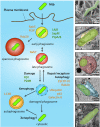
The space: localisation of Mtb within human cells. Once Mtb is phagocytosed by macrophages, Mtb will reside in early phagosomes and inhibition of phagosome-lysosome fusion is central to the survival of Mtb. Mtb uses different strategies to interfere with phagosome maturation. Some Mtb-containing phagosomes may fuse with late endosomes resulting in some mycobacteria localised in late endosomes. On the other hand, Mtb may disrupt the phagosomal membrane and escape to the cytosol. In the example shown, membrane damage is partially observed although artefacts from chemical fixation cannot be excluded. These different stages are not stable but highly dynamic. The panel on the right shows representative EM images of Mtb-infected human macrophages illustrating the different cellular compartments where Mtb can localise/reside within host cells. Host molecules associated with the different localisation stages are shown in orange and Mtb factors implicated in the process shown in green.
Figure 3.
Figure 3.
The time: spatiotemporal dynamics of Mtb interactions with host cell organelles. At least four different populations of intracellular Mtb (early phagosome, late phagosome, damaged phagosome and free in the cytosol) are localised in different environments and will interact dynamically with host cell organelles such as vesicles from the endocytic pathway (e.g. early, recycling and late endosomes); the autophagy pathway, endoplasmic reticulum, post-Golgi vesicles, the extensive network of mitochondria, lipid droplets and peroxisomes. Because of the dynamic nature of these Mtb populations, their interactions with host organelles will be different and likely regulated in a spatiotemporal manne by the bacteria and the host cell. Distinct molecular players involved in these interactions are shown. Known host factors are shown in black and Mtb factors in green.
Figure 4.
Figure 4.
Human macrophage systems to study Mtb-macrophage interactions in vitro. A commonly used human macrophage source is derived from monocytic leukaemia cell lines, like THP-1 and U937 cells that require a ‘differentiation’ step typically achieved with phorbol esters and/or Vitamin D treatment. An alternative source is based on lineage conversion from malignant B-lineage cells to monocytes/macrophages that is caused by the inducible nuclear translocation of a C/EBPα transgene, BLaER1 cells. Although trans-differentiated BLaER1-monocytes transcriptome is highly similar to that of their primary monocytes, if this method can recapitulate the complete functional properties of human primary macrophages requires further validation. Alveolar macrophages can be obtained in limited numbers from broncholavelolar lavage or lung biopsies. Another widely used method is the isolation of monocytes from peripheral blood that are subsequently differentiated using recombinant differentiation factors such as macrophage colony stimulating factor (M-CSF) or granulocyte macrophage colony stimulating factor (GM-CSF). With advances in our understanding of stem cell biology, the use of iPSDM. It involves a stepwise differentiation programme that includes haemogenic endothelial specification achieved and maintained via the introduction of bone morphogenetic protein 4(BMP4), fibroblast growth factor 2 (FGF2) and vascular endothelial growth factor (VEGF). Further differentiation requires signalling from the activin–Nodal and WNT pathways that is achieved through endogenous signalling via the formation of embryoid bodies (EBs). When EBs form, they are transferred to an adherent surface and exposed to cytokines, such as M-CSF and IL-3 to promote the generation of monocytes, which will be finally differentiated into macrophages.
Figure 5.
Figure 5.
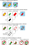
Different fluorescent- based approaches to study the intracellular localisation of Mtb. A, One of the most widely used strategies to study bacteria localisation is based on a biased judgement to determine if a particular intracellular marker (shown in red) co-localise with bacteria (shown in green). B, Alternatively, in cases where the cellular markers co-localise almost completely with bacteria, a more quantitative and unbiased method can be employed using a masking strategy to select bacteria and applying this selection to the cellular marker channel/image to determine the pixel number and intensity associated with bacteria. C, A variant of this unbiased method can also be applied when the intracellular marker is associated with the bacteria membrane but it does not cover all the surface. In this case, is necessary to dilate the original bacteria mask. The basic effect of this operator (on a binary image) is to gradually enlarge the boundaries of regions of foreground pixels. Then, the bacteria pixels will be subtracted and the resulting mask will be used to quantify the corresponding pixels in the cellular marker image/channel. D, Examples where unbiased methods are not suitable for quantification and positive or negative co-localisation is difficult to determine since the distribution of the intracellular marker is asymmetric or it only partially covers the bacteria surface. In these cases, artificial intelligence represents a promising methodological advance.
Figure 6.
Figure 6.
Measuring Mtb viability in host cells after infection. A, Currently, the conventional method to evaluate viability of Mtb relies on bacterial CFU enumeration on agar plates. This is a time-consuming approach due to the slow growth rate of Mtb that takes about 3–6 weeks to observe visible colonies on agar plates. One of its main disadvantages of this method is that clumps of bacteria cells can be miscounted as single colonies. B,The graph illustrates a genetic approach where Mtb constitutively express mCherry (red) and express GFP (green) using an inducible TetON promoter. After tetracycline induction ‘live’ bacteria express both GFP (green) and mCherry (red) fluorescence, while ‘dead’ bacteria are only mCherry positive (red). C, A similar strategy to (B) where the Mtb reporter strains are constructed to constitutively express mCherry (smyc′::mCherry) that enables the visualization of all bacteria combined with the expression of GFP that is regulated by promoters that respond to environmental cues (for example, pH). D, Dual-targeting fluorogenic probes (CDG-DNBs) containing a BlaC-sensing unit, a caged fluorescent reporter, and a DprE1-binding unit for signal trapping. CDG-DNBs pass the Mtb cell wall through porins, BlaC and DprE1 enzymes located in the peptidoglycan layer and at the outer membrane will react with the probes. Then, BlaC will hydrolyze the lactam ring to activate the fluorophore, and DprE1 will covalently bind the anchor unit for fluorescence immobilization. The combined actions of BlaC and DprE1 would enable fluorescent labelling of single Mtb. Bacteria without any BlaC and/or DprE1 activity would not fluorescently label due to the lack of fluorescence activation (no BlaC) or signal retention (no DprE1) in cells. E, A FITC-trehalose probe that exploits the processing by Mtb Ag85 enzymes is specifically incorporated into Mtb growing in vitro and within macrophages.
Figure 7.
Figure 7.
Measuring Mtb intracellular replication. A, One approach to follow Mtb replication involves the quantification of fluorescent Mtb strains through time at single cell level in fixed cells. Although this method can be adapted to high-content imaging, it does not take into account cellular heterogeneity and does not allow to fully distinguish live from dead bacteria. B, Shows the Luciferin-luciferase reporter. This system is useful for studying cell populations but does not allow for single-cell analysis. C, A more time-consuming approach that it does allow spatiotemporal resolution is to evaluate intracellular bacteria replication by live cell imaging. D, Shows genetically encoded fluorescent reporter in combination with quantitative time-lapse microscopy. It uses a destabilized variant of green fluorescent protein (GFPdes) with a plasmid expressing a stable red fluorescent protein (DsRed2) from a constitutive promoter, as internal control. In this example, stationary-phase populations exhibit a drop in GFPdes expression followed by the appearance of a subset of non-replicating bacteria displaying high levels of fluorescence. E, Another replication reporter uses a SSB that is fused to GFP, present on a replicating plasmid that contains a constitutively expressed mCherry. The SSB-GFP reporter defined cell cycle timing, with SSB-GFP foci present for periods of DNA replication and disappearing when the bacteria stop growing or it is during stationary phase.
Figure 8.
Figure 8.
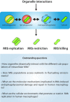
Spatiotemporal regulation of Mtb replication or control. In host cells, Mtb initially resides inside phagosomes where bacteria subvert phagosomal function. Some Mtb will effectively be targeted to late phagosomes where bacteria will be exposed to an acidic and proteolytic environment. At some point during the infection cycle, Mtb will damage phagosomes and access the cytosol. During these events, bacteria-containing compartments and cytosolic bacteria is actively interacting with a plethora of host cell organelles. The interactions between the different Mtb populations and host cell organelles will determine if Mtb replicates, or its growth is restricted or eventually eliminated by the host cell. Outstanding questions are shown emphasizing central themes in the cell biology of Mtb-host cell interactions requiring further research.

References

    1. Alonso S, Pethe K, Russell DGet al. .. Lysosomal killing of Mycobacterium mediated by ubiquitin-derived peptides is enhanced by autophagy. Proc Natl Acad Sci USA. 2007;104:6031–6. - PMC - PubMed
    1. Andreu N, Fletcher T, Krishnan Net al. .. Rapid measurement of antituberculosis drug activity in vitro and in macrophages using bioluminescence. J Antimicrob Chemother. 2012;67:404–14. - PMC - PubMed
    1. Andreu N, Zelmer A, Fletcher Tet al. .. Optimisation of bioluminescent reporters for use with mycobacteria. PLoS One. 2010;5:e10777. - PMC - PubMed
    1. Anes E, Kuhnel MP, Bos Eet al. .. Selected lipids activate phagosome actin assembly and maturation resulting in killing of pathogenic mycobacteria. Nat Cell Biol. 2003;5:793–802. - PubMed
    1. Armstrong JA, Hart PD. Phagosome-lysosome interactions in cultured macrophages infected with virulent tubercle bacilli. Reversal of the usual nonfusion pattern and observations on bacterial survival. J Exp Med. 1975;142:1–16. - PMC - PubMed

Publication types