Review
. 2019 Jun 4:21:443-468.
doi: 10.1146/annurev-bioeng-060418-052139. Epub 2019 Mar 27.

The Driving Force: Nuclear Mechanotransduction in Cellular Function, Fate, and Disease

Affiliations
Review

The Driving Force: Nuclear Mechanotransduction in Cellular Function, Fate, and Disease

Melanie Maurer et al. Annu Rev Biomed Eng. .

Abstract

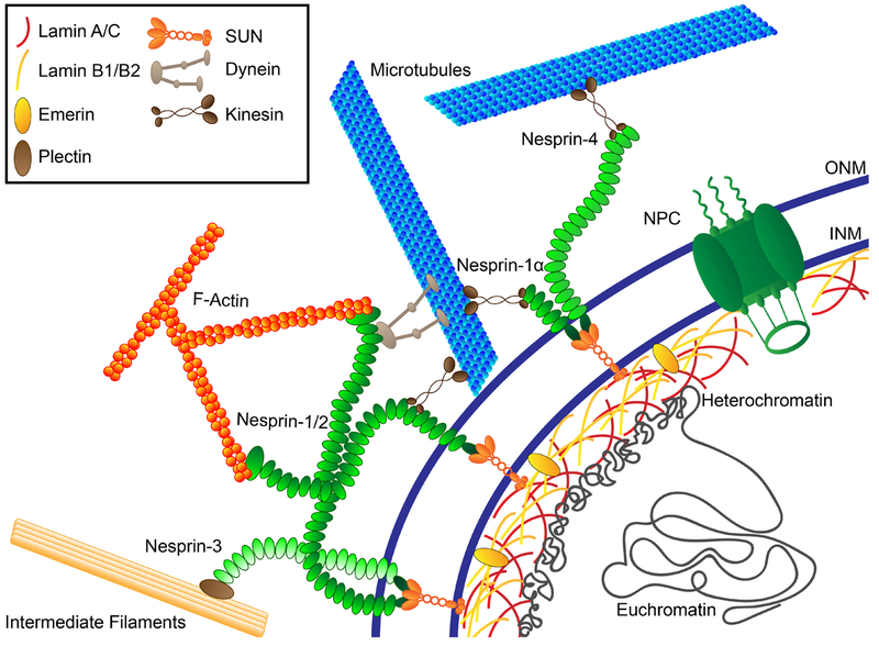
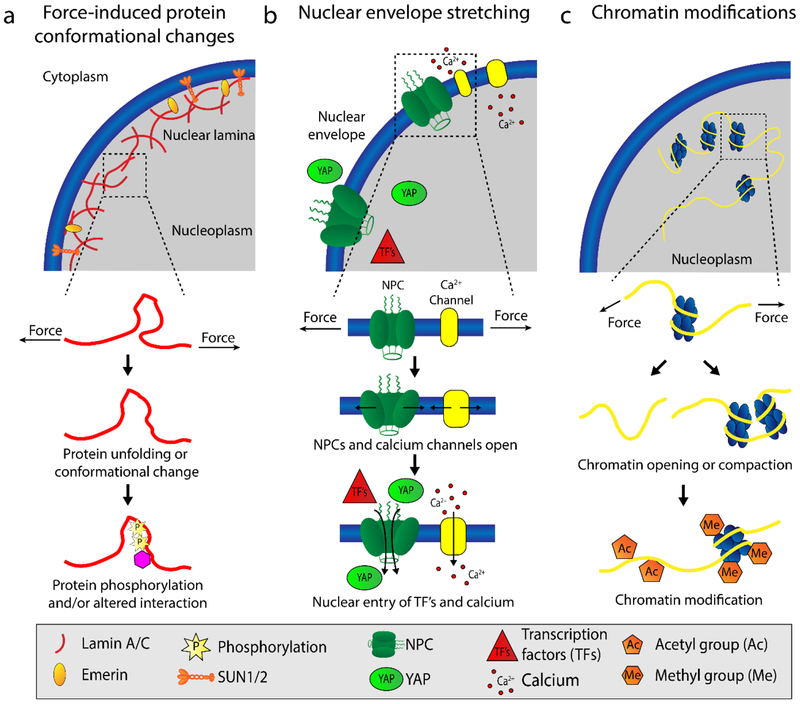
Cellular behavior is continuously affected by microenvironmental forces through the process of mechanotransduction, in which mechanical stimuli are rapidly converted to biochemical responses. Mounting evidence suggests that the nucleus itself is a mechanoresponsive element, reacting to cytoskeletal forces and mediating downstream biochemical responses. The nucleus responds through a host of mechanisms, including partial unfolding, conformational changes, and phosphorylation of nuclear envelope proteins; modulation of nuclear import/export; and altered chromatin organization, resulting in transcriptional changes. It is unclear which of these events present direct mechanotransduction processes and which are downstream of other mechanotransduction pathways. We critically review and discuss the current evidence for nuclear mechanotransduction, particularly in the context of stem cell fate, a largely unexplored topic, and in disease, where an improved understanding of nuclear mechanotransduction is beginning to open new treatment avenues. Finally, we discuss innovative technological developments that will allow outstanding questions in the rapidly growing field of nuclear mechanotransduction to be answered.

Keywords: LINC complex; lamin; laminopathies; mechanotransduction; nuclear mechanics; stem cells.


Figures

Figure 1
Figure 1
Constituents of the nucleus and nuclear envelope involved in mechanotransduction. The LINC (linker of the nucleoskeleton and cytoskeleton) complex—nesprins at the outer nuclear membrane (ONM) and SUN-domain proteins at the inner nuclear membrane (INM)—spans the nuclear envelope, interacting with cytoskeletal filaments and associated proteins and the nuclear lamina to enable force transmission between the cytoskeleton and nuclear interior. Nuclear lamins (A/C and B1/2) form independent yet interacting meshworks underneath the INM and are responsible for maintaining nuclear shape and stiffness. Both A- and B-type lamins interact with nuclear pore complexes (NPCs), chromatin, and various other binding partners at the nuclear envelope and the nuclear interior. NPCs enable molecular transport between the cytoplasm and nucleoplasm.
Figure 2
Figure 2
Proposed mechanisms of nuclear mechanotransduction. (a) Force application to the nucleus can results in conformational changes of nuclear envelope proteins, such as partial unfolding of lamins (11, 71), and phosphorylation of nuclear proteins, including lamins, SUN-domain proteins, and Emerin (10, 11, 56). (b) Nuclear membrane stretch in response to force opens nuclear pore complexes (NPCs) (59, 60) and calcium channels (65, 66) on the cytoplasmic side, thus increasing molecular influx into the nucleoplasm. The increased import of transcription factors (TFs) into the nucleoplasm can alter gene expression (60). (c) Mechanical forces acting on the nucleus can induce chromatin stretching, opening, and compaction, including DNA and histone modifications, that alter accessibility to transcription factors and lead to changes in gene expression (–77) [add reference (82)]. Abbreviation: NPC, nuclear pore complex.
Figure 3
Figure 3
Nuclear mechanics guide stem cell fate and mechanical memory. (a) Stem cells may undergo mechanically induced differentiation in response to matrix mechanical properties, surface structure, and geometry. Nuclear mechanotransduction in response to matrix sensing alters the transcriptional program to ultimately guide downstream lineage commitment and cellular mechanical properties. (b) Substrate stiffness may enable stem cells to exhibit mechanical memory, in which a stiff phenotype is remembered upon transfer to culture on a soft substrate, via nuclear YAP retention and chromatin condensation.
Figure 4
Figure 4
Defective mechanotransduction as a bridge between laminopathy hypotheses. Structural defects (increased nuclear fragility that leads to breakage and cell death) and gene misregulation (altered gene activation and silencing) are the two primary hypothesized mechanisms responsible for the muscle-specific defects in many laminopathies. A third hypothesis—defective nuclear mechanotransduction—synthesizes both the structural disruption and gene misregulation hypotheses, as it can explain how downstream gene misregulation might be a product of nuclear weakness due to disruption of mechanotransduction mechanisms in and on the nucleus.
Figure 5
Figure 5
Creation of engineered muscle tissue constructs for the study of tissue morphology and generated forces. (a) Devices are loaded with a cell (light brown) and extracellular matrix (pink) solution, and (b) cells reorganize and restructure the matrix to form a tissue around elastic pillars. (c) Tissues gradually compact and/or contract as cells elongate, thereby deflecting pillars. The force generated by the engineered tissue constructs can be calculated from the measured pillar deflection and the known material properties of the elastic pillars.

References

    1. Wang N, Tytell JD, Ingber DE. 2009. Mechanotransduction at a distance: mechanically coupling the extracellular matrix with the nucleus. Nat. Rev. Mol. Cell Biol 10:75–82 - PubMed
    1. Doyle AD, Yamada KM. 2016. Mechanosensing via cell–matrix adhesions in 3D microenvironments. Exp. Cell Res 343:60–66 - PMC - PubMed
    1. Cho S, Irianto J, Discher DE. 2017. Mechanosensing by the nucleus: from pathways to scaling relationships. J. Cell Biol 216:305–15 - PMC - PubMed
    1. Jaalouk DE, Lammerding J. 2009. Mechanotransduction gone awry. Nat. Rev. Mol. Cell Biol 10:63–73 - PMC - PubMed
    1. Wang N, Butler JP, Ingber DE. 1993. Mechanotransduction across the cell surface and through the cytoskleton. Science. 260:1124–27 - PubMed

Publication types

LinkOut - more resources