. 2019 Jun;33(6):7647-7666.
doi: 10.1096/fj.201802264R. Epub 2019 Mar 27.

The NMR-based characterization of the FTY720-SET complex reveals an alternative mechanism for the attenuation of the inhibitory SET-PP2A interaction

Affiliations

The NMR-based characterization of the FTY720-SET complex reveals an alternative mechanism for the attenuation of the inhibitory SET-PP2A interaction

Ryan M De Palma et al. FASEB J. 2019 Jun.

Abstract

The su(var)3-9, enhancer of zeste, trithorax (SET)/inhibitor 2 of protein phosphatase 2A (PP2A) oncoprotein binds and inhibits PP2A, composed of various isoforms of scaffolding, regulatory, and catalytic subunits. Targeting SET with a sphingolipid analog drug fingolimod (FTY720) or ceramide leads to the reactivation of tumor suppressor PP2A. However, molecular details of the SET-FTY720 or SET-ceramide, and mechanism of FTY720-dependent PP2A activation, remain unknown. Here, we report the first in solution examination of the SET-FTY720 or SET-ceramide complexes by NMR spectroscopy. FTY720-ceramide binding resulted in chemical shifts of residues residing at the N terminus of SET, preventing its dimerization or oligomerization. This then released SET from PP2ACα, resulting in PP2A activation, while monomeric SET remained associated with the B56γ. Our data also suggest that the PP2A holoenzyme, composed of PP2A-Aβ, PP2A-B56γ, and PP2ACα subunits, is selectively activated in response to the formation of the SET-FTY720 complex in A549 cells. Various PP2A-associated downstream effector proteins in the presence or absence of FTY720 were then identified by stable isotope labeling with amino cells in cell culture, including tumor suppressor nonmuscle myosin IIA. Attenuation of FTY720-SET association by point mutations of residues that are involved in FTY720 binding or dephosphorylation of SET at Serine 171, enhanced SET oligomerization and the formation of the SET-PP2A inhibitory complex, leading to resistance to FTY720-dependent PP2A activation.-De Palma, R. M., Parnham, S. R., Li, Y., Oaks, J. J., Peterson, Y. K., Szulc, Z. M., Roth, B. M., Xing, Y., Ogretmen, B. The NMR-based characterization of the FTY720-SET complex reveals an alternative mechanism for the attenuation of the inhibitory SET-PP2A interaction.

Keywords: ceramide; fingolimod; lipid-protein binding; sphingolipid.


Conflict of interest statement

The authors thank Dr. Aiping Bai and Mr. Jason Pierce for help in ceramide measurements at the Lipidomics Shared Resource facility, Department of Biochemistry and Molecular Biology, Medical University of South Carolina. The authors also thank Mr. Brett Bechtol (Hollings Cancer Center, Medical University of South Carolina) for editorial review and assistance. The authors thank the members of the B.O. laboratory for technical support and helpful discussions. The authors also thank Dr. Do-Sung Kim (Department of Biochemistry and Molecular Biology, Hollings Cancer Center, Medical University of South Carolina) for help and assistance in our experimental procedures. This work is supported by research funding from the U.S. National Institutes of Health (NIH) National Cancer Institute (CA088932, CA173687, CA214461, and P01 CA203628, to B.O.), and the NIH National Institute of Dental and Craniofacial Research (DE016572, to B.O.). The core facilities utilized were constructed using support from NIH National Center for Research Resources (C06 RR015455), Hollings Cancer Center Support Grant (P30 CA138313), or Center of Biomedical Research Excellence (CoBRE) in Lipidomics and Pathobiology (P30 GM103339). B.O. is the founder and Chief Scientific Officer of Lipo-Immuno Tech, LLC., and Z.M.S. is the director of synthetic chemistry of the Lipo-Immuno Tech. The authors declare no conflicts of interest.

Figures
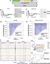
Figure 1
Figure 1
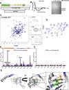
C6P binds SET in an extended conformation. A) Diagram representation on recombinant SET proteins used in this study. B) CD spectra and Coomassie stained Nd-SETWT. C) CD spectra and Coomassie stained Nd-SETK209D. All CD scans were in triplicate, averaged, and from 195 to 260 nm wavelength range at 20°C. D) ITC of 0.05 mM C6P titrated into 300 μl of 0.021 mM Nd-WT-SET. E) ITC of 0.05 mM C6P titrated into 300 μl of 0.021 K209D Nd-SET. Compiled ITC data presented as averages of triplicate experiments ± sem. F) STD-NMR saturation buildup of 0.5 mM C6P. G) STD-NMR buildup curves were calculated using STD-AF isotherm. H) Proposed model of ceramides binding in an extended conformation.
Figure 2
Figure 2
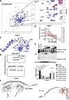
d-e-C18-ceramide, but not its enantiomer l-e-C18-ceramide, binds to SET. A) Graphic representation on recombinant SET proteins used in this study. B) CD spectra and Coomassie stained NdCd-SETWT. C) [1H-15N] TROSY spectra of 0.9 mM NdCd-SET. Expansion of spectral regions showing specific perturbed residues upon binding with d-e-C18-ceramide. D) Interaction diagram of d-e-C18-ceramide binding suggests that one of the prominent binding sites includes the E111, V112, and Y127. All CSP titrations were performed in triplicate. E) CSP of NdCd-SET induced by 3 μM (black bars), 7 μM (red bars), and 10 μM D-e-C18-ceramide (blue bars) as a function of assigned residue. Red line is calculated as 3σ, and any value >3σ is considered a significant shift. F) CSP of NdCd-SET induced by 3 μM (black bars), 7 μM (red bars), and 10 μM l-e-C18 ceramide (blue bars) as a function of assigned residue. Red line is calculated as 3σ, and any value >3σ is considered a significant shift. G) Model of d-e-C18-ceramide–bound SET. Residues colored blue and green underwent a significant chemical shift during NMR titration analysis.
Figure 3
Figure 3
FTY720 binds SET similarly as d-e-C18 ceramide. A) Overlaid [1H-1H] total correlational spectroscopy spectra of FTY720. Quantitation of line broadening as a function of peak volume and increasing concentration of Nd-SETWT (0–50 μM). Chemical structure of functional groups on FTY720 affected by the addition of Nd-SET. B) CSP of NdCd-SET induced by 3 μM (black bars), 7 μM (red bars), and 10 μM FTY720 (blue bars) as a function of assigned residue. CSP titrations were performed in triplicate. Red line is calculated as 3σ, and any value >3σ is considered a significant shift. C) Model of FTY720 bound SET. Residues colored red underwent a significant chemical shift during NMR titration analysis. D) Association between endogenous SET, FLAG-SETWT, FLAG-SETE111A, and FLAG-SETR71A mutants and PP2AC ± 5 μM FTY720 was monitored by PLA using antibodies against SET, FLAG, or PP2AC. SET mutants were generated from CSP and modeling. Representative images of n ≥ 3 independent experiments per group. Scale bar, 100 μm. E) Quantitation of PLA for SET mutants designed to limit response to FTY720 treatment. Data are means ± sem of n ≥3 independent experiments per group, analyzed by 2-way ANOVA with Tukey’s post hoc test. *P < 0.05, ***P < 0.001.
Figure 4
Figure 4
Lipid binding may alter SET dimerization domain to prevent oligomerization. A) Spectrum of NdCd highlights peaks that undergo line broadening upon the addition of FTY720. These peaks correspond to residues in the dimerization domain of SET. Blowout highlights broadened peaks (red) at 10 μM FTY720. B) Quantitation of the total change in peak height of assigned residues in the dimerization domain. Data are means from triplicate experiments. C) Comparison of NdCd-SETWT models in the absence (white) and presence (blue) of d-e-C18 ceramide illustrating predicted structural alterations in the N-terminal helix of SET. Model focuses on region between I42 and P79. D) DOSY spectra and diffusion coefficients ± 10 μM FTY720 were analyzed with DOSY ToolBox 2.7 (53). E) GA cross-linking of SET oligomers in response to 3 h of treatment with 1–5 μM d-e-C18 or l-e-C18-ceramides. Representative blot of n = 3 individual experiments. N.s., not significant. Data were analyzed by Student’s t test. **P < 0.01, ***P < 0.001. F) Working model illustrating the prevention of SET oligomerization due to binding with FTY720.
Figure 5
Figure 5
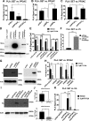
SET interacts with specific PP2A holoenzyme. A–C) Association between endogenous SET and PP2AC in shPP2AA⍺ and shPP2AAβ (A) knockdown cells, interaction between endogenous SET and PP2AC in shB56γ and shB56δ (B) knockdown cells, and association between endogenous SET and PP2AC in shPP2AC⍺ and shPP2ACβ (C) knockdown cells was assessed by PLA. Data are means ± sem of n = 3 independent experiments, analyzed by Student’s t test. *P < 0.05, **P < 0.01. D) Western blot showing successful reconstitution of expression for V5-PP2AA⍺ and V5-PP2AAβ. E) Interaction between endogenous SET and PP2AC in shPP2AA⍺ and shPP2AAβ stable knockdown cells with rescued expression (V5-PP2AA⍺ and V5-PP2AAβ was assessed by PLA using antibodies against SET and PP2AC). Quantitation of PLA. Data are means ± sem of n = 3 independent experiments, analyzed by Student’s t test. ***P < 0.001, ****P < 0.0001. F) Association between endogenous SET and V5-PP2AA⍺ and V5-PP2Aaβ in shPP2AA⍺ and shPP2AAβ stable knockdown cells using antibodies against SET and V5. Quantitation of PLA. Data are means ± sem of n = 3 independent experiments, analyzed by Student’s t test. ****P < 0.0001. G) Western blot showing successful reconstitution of expression for V5-B56γ and V5-B56δ. H) Interaction between endogenous SET and PP2AC in shB56γ and shB56δ stable knockdown cells with rescued expression (V5-B56γ and V5-B56δ) was assessed by PLA using antibodies against SET and PP2AC. Quantitation of PLA. Data are means ± sem of n = 3 independent experiments, analyzed by Student’s t test. *P < 0.05, **P < 0.01, ***P < 0.001. I) Western blot showing successful reconstitution of expression for HA-PP2AC⍺ and HA-PP2ACβ. J) Levels of mRNA showing knockdown of PP2AC⍺ and PP2ACβ obtained by quantitative RT-PCR. K) Association between endogenous SET and HA-tagged PP2AC in shPP2AC⍺ and shPP2ACβ stable knockdown cells with rescued expression (shPP2AC⍺ + HA-PP2AC⍺ and shPP2ACβ + HA-PP2Acβ) was assessed by PLA using antibodies against SET and HA epitope. Quantitation of PLA. Data are means ± sem of n = 3 independent experiments, analyzed by Student’s t test. ****P < 0.0001.
Figure 6
Figure 6
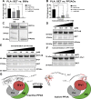
Reconstitution of PP2ACα and B56γ expression restores cellular response to FTY720. A) Interaction between endogenous SET and B56γ ± 5 μM FTY720, 5 μM d-e-C18 ceramide, or 1 μM OP449 treatment for 3 h assessed by PLA using antibodies against SET and B56γ. Quantitation of PLA experiments. Error bars are sem of n ≥3 independent experiments per group. Data are means ± sem of n = 3 independent experiments per group, analyzed by 2-way ANOVA with Tukey’s post hoc test. *P < 0.05. B) Association between endogenous SET and PP2AC X ± 5 μM FTY720, 5 μM d-e-C18 ceramide, or 1 μM OP449 treatment for 3 h assessed by PLA using antibodies against SET and PP2AC. Quantitation of PLA experiments. Error bars are sem of n ≥3 independent experiments per group. Data are means ± sem of n ≥3 independent experiments per group, analyzed by 2-way ANOVA with Tukey’s post hoc test. *P < 0.05, **P < 0.01, ***P < 0.001. C) Pull-down assay with either GST-tagged PP2A core enzyme (AβCα or AαCα) and SET. D) Pull-down assay with either GST-tagged PP2A core enzyme (AβCα) and SET with increasing (0–50 μM) FTY720. E) Pull-down assay with either GST-tagged PP2A holoenzyme (AβCαBγ1) and SET with increasing (0–100 μM) FTY720. F) Pull-down assay with either GST-tagged Bγ1 and SET with increasing (0–50 μM) FTY720. G) SET oligomerization is essential for PP2A inhibition. Lipid binding prevents SET oligomerization resulting in active PP2A. Additionally, SET remains with FTY720-activated PP2A by maintaining contact with the B56γ subunit as a possible regulatory mechanism.
Figure 7
Figure 7
Myosin IIa is a potential target of FTY720-activated PP2A. A) Validation of PP2AC/myosin IIa association observed in SILAC data by coimmunoprecipitation. Exogenous expression of HA-PP2ACαWT in A549 cells using magnetic HA-conjugated beads ± treatment of 20 μM FTY720 for 2 h. B) Association between PP2AC and myosin IIa in shSCR and shB56γ stable knockdown cell lines ± treatment of 5 μM FTY720 for 3 h by PLA using antibodies against PP2AC and myosin IIa. Quantitation of PLA experiments. Data are means ± sem of n = 3 independent experiments, analyzed by 2-way ANOVA with Tukey’s post hoc test. *P < 0.05, **P < 0.01. C) PP2A activity assay in shSCR and shB56γ stable knockdown cell lines ± treatment of 5 μM FTY720 for 16 h using purified rabbit MYH9 as a substrate. Data are means ± sd of n = 3 independent experiments analyzed by 2-way ANOVA with Tukey’s post hoc test. *P < 0.05, ***P < 0.001. D) Western blot of PP2A-p-Tyr307 on PP2AC, normalized to β-actin in shScr and shB56γ stable cell lines. E) PP2A activity assay in A549 cells transiently overexpressing WT, R71A, or E111A FLAG-SET ± treatment of 5 μM FTY720 for 16 h using purified rabbit MYH9 as a substrate. Data are means ± sd of n = 3 independent experiments analyzed by 2-way ANOVA with Tukey’s post hoc test. **P < 0.01, ***P < 0.001. F) Western blot of p-Tyr307 on PP2AC, normalized to β-actin. G) Quantification of PP2A-p-Tyr307 blots. Representative blot of n = 3 individual experiments. Blots were quantified with ImageJ. Data are means normalized to actin ± sd and analyzed by 2-way ANOVA with Tukey’s post hoc test. *P < 0.05, **P < 0.01. H) Validation of p-Tyr307 antibody through exogenous expression of HA-PP2ACαWT or nonphosphorylatable mutant HA-PP2ACαY307A in A549 cells. Input of cellular extracts is 10% of samples for each IP. I) Coimmunoprecipitation of HA-PP2ACαWT or HA-PP2ACαY307A using magnetic HA-conjugated beads 72 h post-transfection.
Figure 8
Figure 8
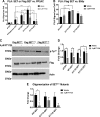
Phosphorylation of SET influences PP2A activity. A) Association between endogenous SET, FLAG-SETWT, FLAG-SETS171A, and FLAG-SETS171E, mutants, and PP2AC ± 5 μM FTY720 with a pCDH empty vector control measured by PLA using antibodies against SET, FLAG, and PP2AC. Data are means ± sem of n = 3 independent experiments, analyzed by 2-way ANOVA with Tukey’s post hoc test. *P < 0.05, **P < 0.01, ****P < 0.0001. B) Association between FLAG-SETS171A and FLAG-SETS171E and B56γ ± 5 μM FTY720 assessed by PLA using antibodies against FLAG and B56γ. Data are means ± sem of n = 3 independent experiments analyzed by 2-way ANOVA with Tukey’s post hoc test. *P < 0.05, **P < 0.01. C) Western blot of PP2A-p-Tyr307 of PP2AC as a marker of activity normalized to β-actin and FLAG expression. Representative blot of n = 3 individual experiments. D) Blots were quantified with ImageJ. Data are means normalized to actin and FLAG ± sd and analyzed by 2-way ANOVA with Tukey’s post hoc test. *P < 0.05. E) Quantification of GA cross-linking of FLAG-SETWT, FLAG-SETS171A, and FLAG-SETS171E oligomers in response to 3-h treatment with 5 μM FTY720. Data are means ± sd (n = 3 independent experiments) analyzed by 2-way ANOVA with Tukey’s post hoc test. *P < 0.05.

References

    1. Janssens V., Goris J. (2001) Protein phosphatase 2A: a highly regulated family of serine/threonine phosphatases implicated in cell growth and signalling. Biochem. J. 353, 417–439 - PMC - PubMed
    1. Virshup D. M. (2000) Protein phosphatase 2A: a panoply of enzymes. Curr. Opin. Cell Biol. 12, 180–185 - PubMed
    1. Herzog F., Kahraman A., Boehringer D., Mak R., Bracher A., Walzthoeni T., Leitner A., Beck M., Hartl F.-U., Ban N., Malmström L., Aebersold R. (2012) Structural probing of a protein phosphatase 2A network by chemical cross-linking and mass spectrometry. Science 337, 1348–1352 - PubMed
    1. Saydam G., Aydin H. H., Sahin F., Selvi N., Oktem G., Terzioglu E., Buyukkececi F., Omay S. B. (2003) Involvement of protein phosphatase 2A in interferon-alpha-2b-induced apoptosis in K562 human chronic myelogenous leukaemia cells. Leuk. Res. 27, 709–717 - PubMed
    1. Bialojan C., Takai A. (1988) Inhibitory effect of a marine-sponge toxin, okadaic acid, on protein phosphatases. Specificity and kinetics. Biochem. J. 256, 283–290 - PMC - PubMed

Publication types

MeSH terms

Substances

LinkOut - more resources