

Clinical Implementation of a Focused MRI Protocol for Hepatic Fat and Iron Quantification
- PMID: 30917020
- PMCID: PMC6764903
- DOI: 10.2214/AJR.18.20947
Clinical Implementation of a Focused MRI Protocol for Hepatic Fat and Iron Quantification
Abstract
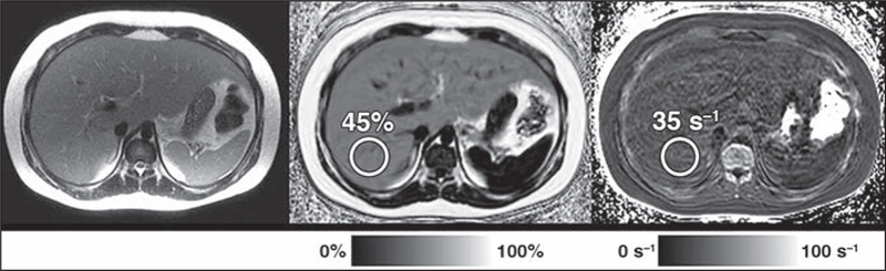
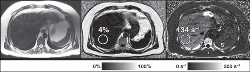
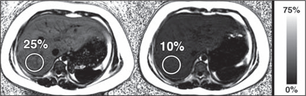
OBJECTIVE. The purpose of this article is to describe our institutional experience with the clinical implementation of a novel focused rapid chemical shift-encoded MRI protocol specifically intended to detect and quantify hepatic steatosis and iron overload, highlighting usage statistics and issues related to cost. CONCLUSION. Focused MRI examinations for specific clinical indications, such as this protocol for detection and quantification of hepatic steatosis and iron overload, are feasible in a busy clinical practice and add value for patients and referring providers.
Keywords: MRI; R2*; chemical shift–encoded MRI; focused protocol; hepatic steatosis; iron overload; limited examination; liver; proton density fat fraction; rapid examination.
Figures
References
-
- Ahmed A, Wong RJ, Harrison SA. Nonalcoholic fatty liver disease review: diagnosis, treatment, and outcomes. Clin Gastroenterol Hepatol 2015; 13:2062–2070 - PubMed
-
- Schwimmer JB, Deutsch R, Kahen T, Lavine JE, Stanley C, Behling C. Prevalence of fatty liver in children and adolescents. Pediatrics 2006; 118:1388–1393 - PubMed
-
- Gramlich T, Kleiner DE, McCullough AJ, Matteoni CA, Boparai N, Younossi ZM. Pathologic features associated with fibrosis in nonalcoholic fatty liver disease. Hum Pathol 2004; 35:196–199 - PubMed
-
- Matteoni CA, Younossi ZM, Gramlich T, Boparai N, Liu YC, McCullough AJ. Nonalcoholic fatty liver disease: a spectrum of clinical and pathological severity. Gastroenterology 1999; 116:1413–1419 - PubMed
-
- Adams LA, Lymp JF, St Sauver J, et al. The natural history of nonalcoholic fatty liver disease: a population-based cohort study. Gastroenterology 2005; 129:113–121 - PubMed
Grants and funding
LinkOut - more resources
Full Text Sources
