. 2019 Sep 1;27(5):466-473.
doi: 10.4062/biomolther.2019.004.

Decursinol Angelate Ameliorates Dextran Sodium Sulfate-Induced Colitis by Modulating Type 17 Helper T Cell Responses

Affiliations

Decursinol Angelate Ameliorates Dextran Sodium Sulfate-Induced Colitis by Modulating Type 17 Helper T Cell Responses

Bikash Thapa et al. Biomol Ther (Seoul). .

Erratum in

Abstract

Angelica gigas has been used as a Korean traditional medicine for pain relief and gynecological health. Although the extracts are reported to have an anti-inflammatory property, the bioactive compounds of the herbal plant and the effect on T cell responses are unclear. In this study, we identified decursinol angelate (DA) as an immunomodulatory ingredient of A. gigas and demonstrated its suppressive effect on type 17 helper T (Th17) cell responses. Helper T cell culture experiments revealed that DA impeded the differentiation of Th17 cells and IL-17 production without affecting the survival and proliferation of CD4 T cells. By using a dextran sodium sulfate (DSS)-induced colitis model, we determined the therapeutic potential of DA for the treatment of ulcerative colitis. DA treatment attenuated the severity of colitis including a reduction in weight loss, colon shortening, and protection from colonic tissue damage induced by DSS administration. Intriguingly, Th17 cells concurrently with neutrophils in the colitis tissues were significantly decreased by the DA treatment. Overall, our experimental evidence reveals for the first time that DA is an anti-inflammatory compound to modulate inflammatory T cells, and suggests DA as a potential therapeutic agent to manage inflammatory conditions associated with Th17 cell responses.

Keywords: Angelica gigas; Decursinol angelate; IL-17; Type 17 helper T cell; Ulcerative colitis.


Conflict of interest statement

CONFLICT OF INTEREST

We wish to confirm that there are no known conflicts of interest associated with this publication and there has been no significant financial support for this work that could have influenced its outcome.

Figures

Fig. 1.
Fig. 1.
Decursinol angelate is an immunomodulatory ingredient of A. gigas. (A–C) Lymph node cells isolated from OT-II mice were treated with the chemical components of A. gigas and activated with ova peptide and culture for 5 days in the presence of IL-2. The culture supernatant was analyzed by CBA. Data represent the mean (± SD) cytokine levels obtained from three independent experiments: *p<0.05, **p<0.01.
Fig. 2.
Fig. 2.
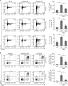
DA suppresses Th17 differentiation and function. CD4+ T cells were activated and cultured in the presence of DA under Th17 polarizing condition for 5 days. (A) Cells were analyzed by intracellular cytokine staining. Shown are representative FACS profiles in the live CD4 T cell gate: inset numbers indicate the mean (± SD) percentages of IL-17+ cells obtained from three independent experiments. (B) Cells were restimulated with anti-CD3e and anti-CD28 for 24 h, and the culture supernatants were analyzed by CBA. (C, D) Th17 cells were cultured as in (A) and analyzed by quantitative RT-PCR. Concentrations of the indicated mRNAs were normalized to those of Actb, and the means (± SD) of the relative levels from three biological replicates are shown: *p<0.05, **p<0.01.
Fig. 3.
Fig. 3.
Effect of DA on survival and proliferation of CD T cells. CD4 T cells were activated with anti-CD3 and anti-CD28 for 3 days in the presence or absence of DA. Cell viability (A) was determine with the Ez-Cytox kit, and apoptosis (B) was measured by Annexin-V staining. The data represent the means (± SD) of three independent experiments. (C) CD4 T cells were cultured as in (A) and analyzed by flow cytometry after a pulse with EdU. Shown are representative histograms in the live CD4 T cell gates and the means (± SD) of EdU+ cells averaging three independent experiments.
Fig. 4.
Fig. 4.
DA ameliorates DSS-induced colitis in mice. (A) Schematic diagram of experimental procedure. (B) Body weight was measured daily and represented as a mean (± SEM) percentage of body weight. (C) Disease activity index (DAI) was scored as describe in “Materials and Methods”. Shown are the means (± SEM) of DAI 9 d after starting DSS administration. Results are obtained from two separate replicate experiments, each involving six mice. (D) At day 9, mice were euthanized, and the colons were isolated and photographed. Colon length was measured. (E) IL-6 level in the plasma at day 9 was analyzed by CBA. (F) Colon were stained with hematoxylin and eosin (H&E). Shown are the representative H&E-stained sections and means (± SD) of the histological scores. *p<0.05, **p<0.01.
Fig. 5.
Fig. 5.
DA attenuates the activation of Th17 cells and the recruitment of neutrophils in the colitis tissues. (A) Mice were administered with DSS as in Fig. 4, and leukocytes isolate from the mesenteric lymph node (MLN), colonic intraepithelium (colonic IE) and lamina propria (colonic LP) were analyzed by intracellular cytokine staining. Shown are the representative FACS profiles with the inset numbers indicating the prevalence of the IL-17+ and/or IFN-γ+ cells in the live CD4 T cell gate. The mean (± SD) percentages of the IL-17A+ cells were calculated from two independent experiments, each involving four mice. (B) Infiltration of granulocyte populations in the colonic tissues were analyzed by flow cytometry. Shown are the representative FACS profiles in the live leukocyte gate and mean (± SD) prevalence of CD11bhi Gr-1hi. *p<0.05, **p<0.01.

References

    1. Allez M, Mayer L. Regulatory T cells: peace keepers in the gut. Inflamm Bowel Dis. 2004;10:666–676. doi: 10.1097/00054725-200409000-00027. - DOI - PubMed
    1. Baumgart DC, Carding SR. Inflammatory bowel disease: cause and immunobiology. Lancet. 2007;369:1627–1640. doi: 10.1016/S0140-6736(07)60750-8. - DOI - PubMed
    1. Boschetti G, Kanjarawi R, Bardel E, Collardeau-Frachon S, Duclaux-Loras R, Moro-Sibilot L, Almeras T, Flourie B, Nancey S, Kaiserlian D. Gut inflammation in mice triggers proliferation and function of mucosal Foxp3+ regulatory T cells but impairs their conversion from CD4+ T cells. J Crohns Colitis. 2017;11:105–117. doi: 10.1093/ecco-jcc/jjw125. - DOI - PubMed
    1. Cho JH, Kwon JE, Cho Y, Kim I, Kang SC. Anti-inflammatory effect of Angelica gigas via heme oxygenase (HO)-1 expression. Nutrients. 2015;7:4862–4874. doi: 10.3390/nu7064862. - DOI - PMC - PubMed
    1. Choi KO, Lee I, Paik SY, Kim DE, Lim JD, Kang WS, Ko S. Ultrafine Angelica gigas powder normalizes ovarian hormone levels and has antiosteoporosis properties in ovariectomized rats: particle size effect. J. Med Food. 2012;15:863–872. doi: 10.1089/jmf.2011.2047. - DOI - PMC - PubMed