. 2019 May 6;216(5):1027-1037.
doi: 10.1084/jem.20181430. Epub 2019 Mar 27.

Aire-expressing ILC3-like cells in the lymph node display potent APC features

Affiliations

Aire-expressing ILC3-like cells in the lymph node display potent APC features

Tomoyoshi Yamano et al. J Exp Med. .

Abstract

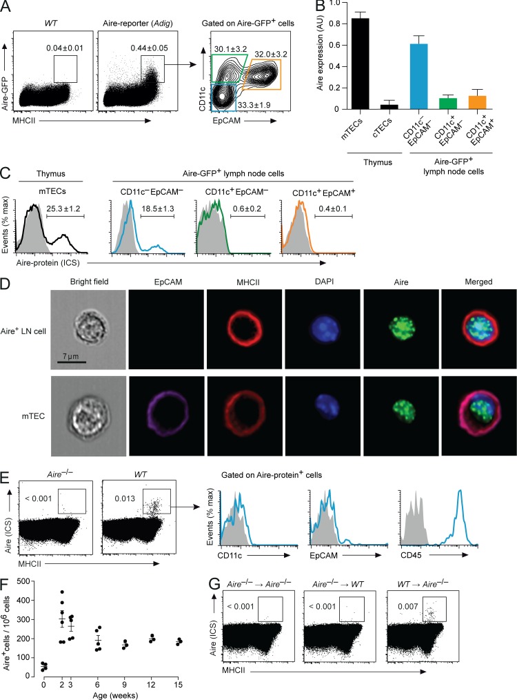
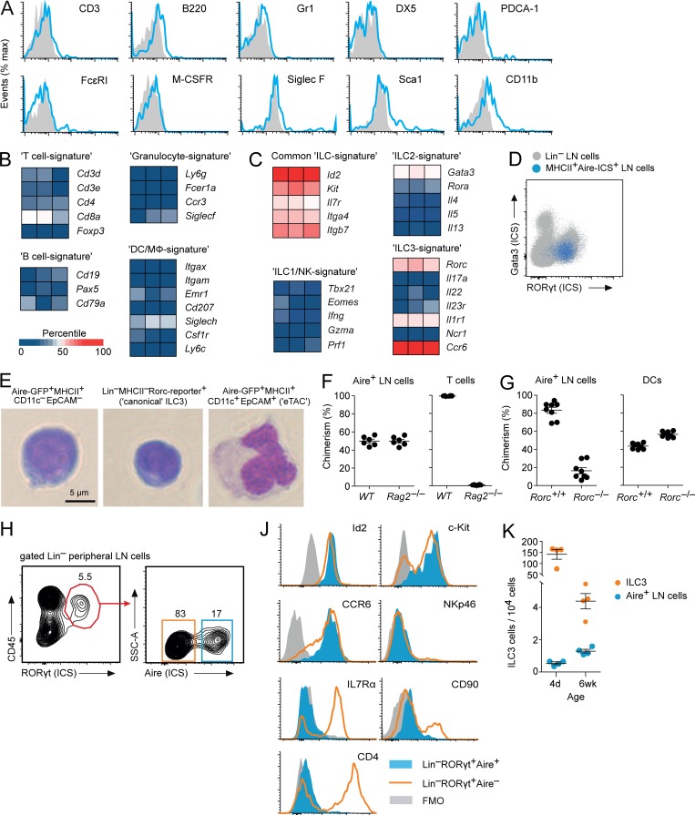
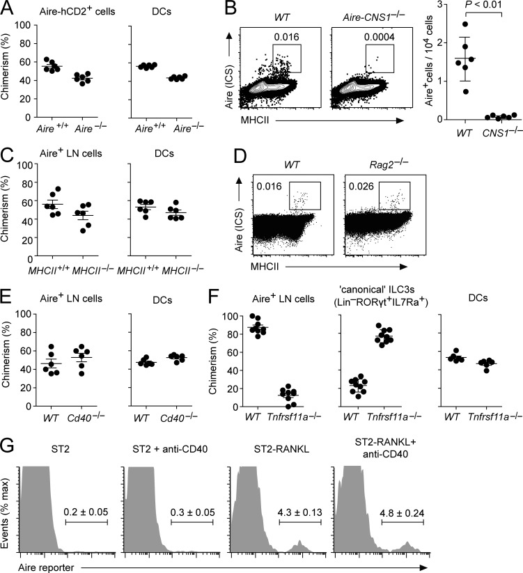
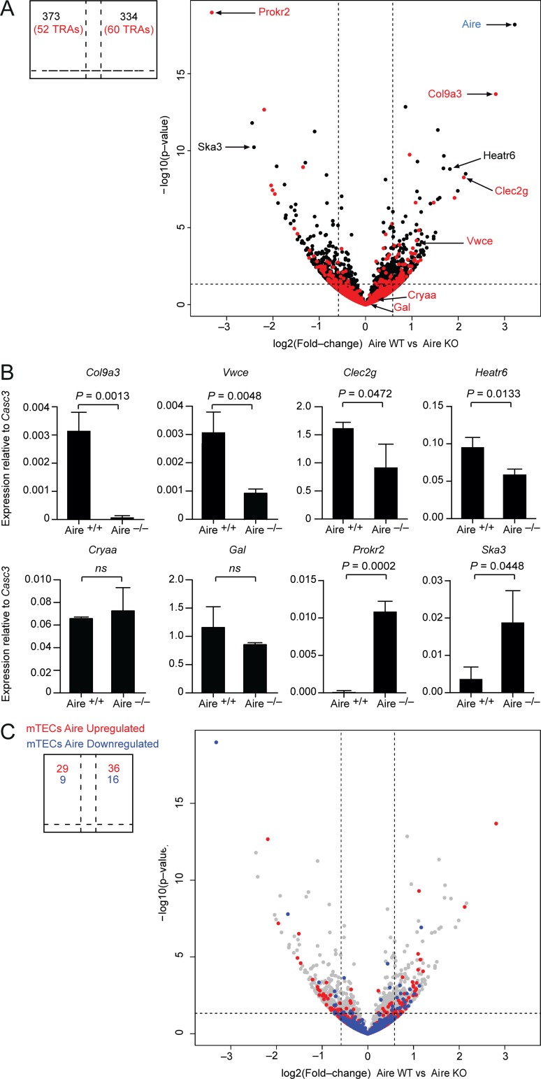
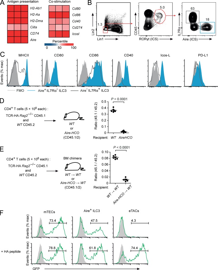
The autoimmune regulator (Aire) serves an essential function for T cell tolerance by promoting the "promiscuous" expression of tissue antigens in thymic epithelial cells. Aire is also detected in rare cells in peripheral lymphoid organs, but the identity of these cells is poorly understood. Here, we report that Aire protein-expressing cells in lymph nodes exhibit typical group 3 innate lymphoid cell (ILC3) characteristics such as lymphoid morphology, absence of "classical" hematopoietic lineage markers, and dependence on RORγt. Aire+ cells are more frequent among lineage-negative RORγt+ cells of peripheral lymph nodes as compared with mucosa-draining lymph nodes, display a unique Aire-dependent transcriptional signature, express high surface levels of MHCII and costimulatory molecules, and efficiently present an endogenously expressed model antigen to CD4+ T cells. These findings define a novel type of ILC3-like cells with potent APC features, suggesting that these cells serve a function in the control of T cell responses.


Figures

Figure 1.
Figure 1.
Phenotype of Aire-expressing cells in LNs. (A) Expression of GFP and MHCII in LN cells from Adig Aire-reporter mice and WT controls and staining for CD11c and EpCAM on gated Aire-GFP+MHCII+ cells (representative of n ≥ 4 each). (B) Aire mRNA in medullary and cortical thymic epithelial cells (mTECs and cTECs, respectively) and in Aire-GFP+ LN cells sorted according to expression of CD11c and EpCAM. Data are mean values ± SEM of triplicates. AU, arbitrary units. (C) ICS for Aire protein in subsets of Aire-GFP+ LN cells. The average frequency ± SEM of Aire-ICS+ cells is indicated (n = 4). (D) Nuclear localization of Aire protein and surface marker expression in Aire-expressing LN cells or mTECs visualized by imaging flow cytometry. (E) ICS for Aire protein and surface expression of MHCII in total LN cells from WT and Aire−/− mice. Histograms on the right show CD11c, EpCAM, and CD45 on gated Aire+MHCII+ cells (representative of n ≥ 5). (F) Number of Aire+ LN cells per 106 total pLN cells in mice of the indicated age (n ≥ 3 each; 0 wk = 4 d old). (G) Aire and MHCII expression in total LN cells from Aire−/−Aire−/−, Aire−/−WT or WTAire−/− BM chimeras (representative of n ≥ 4 each).
Figure 2.
Figure 2.
Aire+ LN cells display ILC3 characteristics. (A) Expression of hematopoietic lineage markers on Aire-GFP+MHCII+CD11cEpCAM LN cells from Adig mice (representative of n ≥ 4). (B) RNA expression of hematopoietic lineage-specific signature genes in Aire+ LN cells. (C) RNA expression of ILC signature genes in enriched Aire+ LN cells. (D) ICS for RORγt and Gata3 in MHCII+Aire+ LN cells (blue dots) back-gated on total Lin (CD3, CD19, B220, Gr-1, CD11c, and CD11b) LN cells (gray dots; representative of n = 3). (E) Morphology of representative Aire-GFP+MHCII+CD11cEpCAM LN cells, “canonical” LN ILC3s (LinMHCIIRORγt+IL7Rα+), and eTACs (Aire-GFP+MHCII+EpCAM+ CD11c+). (F) Contribution of WT and Rag2−/− precursor cells to Aire-ICS+ LN cells or T cells in 1:1 mixed [Rag2−/−: WT] → WT BM-chimeras (n = 6). (G) Contribution of Rorc+/+ and Rorc−/− precursor cells to Aire-ICS+ cells in LN or spleen or to DCs in 1:1 mixed [Rorc+/+: Rorc−/−] → WT BM-chimeras (n = 7). (H) Gating strategy for Lin (Lin1: CD3, CD19, B220, Gr-1; Lin 2: CD11c and CD11b) RORγt+ LN ILC3s and expression of Aire by ICS. (J) Intracellular staining for Id2 protein and surface expression of c-kit, CCR6, NKp46, IL7Rα, CD90, and CD4 in Aire+ (filled blue histogram) and Aire (open orange histogram) LinRORγt+ cells (representative of n = 3). FMO, fluorescence minus one. (K) Number of “canonical” LN ILC3s (orange) and Aire+ LN cells (blue) in 4-d- and 6-wk-old animals (n = 4). Data are mean ± SEM.
Figure 3.
Figure 3.
Molecular and cellular requirements for Aire expression in Aire+ ILC3-like cells. (A) Contribution of Aire+/+ and Aire−/− precursor cells to Aire-reporter+ LN cells or DCs in 1:1 mixed [Aire+/+ Aire-HCO: Aire−/− Aire+/+ Aire-HCO] → WT BM-chimeras (n = 6). (B) Aire (ICS) and MHCII in LN cells from WT controls and Aire-CNS1−/− mice and number of Aire-ICS+MHCII+ cells per 104 LN cells (n = 6 each). (C) Contribution of MHCII+/+ and MHCII−/− precursor cells to Aire+ LN cells or DCs in 1:1 mixed [MHCII+/+: MHCII−/−] → WT BM-chimeras (n = 6). (D) Aire (ICS) and MHCII in LN cells from Rag2−/− mice and WT controls (n ≥ 3 each). (E) Contribution of Cd40+/+ and Cd40−/− precursor cells to Aire+ LN cells or DCs in 1:1 mixed [Cd40+/+: Cd40−/−] → WT BM-chimeras (n = 6). (F) Contribution of Tnfrsf11a+/+ and Tnfrsf11a−/− precursor cells to Aire+ LN cells, “canonical” ILC3s (LinRORγt+IL7Rα+), or DCs in 1:1 mixed [Tnfrsf11a+/+: Tnfrsf11a−/−] → WT fetal liver chimeras (n = 9). (G) Aire-reporter expression in “canonical” IL7Rα+ LN ILC3s after in vitro culture with or without agonistic anti–CD40 antibody or with ST2-RankL cells (representative of n = 3 each). Data are mean ± SEM. Student’s t test was used to calculate P values.
Figure 4.
Figure 4.
Aire orchestrates a distinct genetic program in Aire+ ILC3-like cells. (A) RNA-seq data from LinMHCIIhiCD80+ cells from Aire+/+ and Aire−/− mice. Genes encoding for TRAs are colored in red. Fold-change cutoff: 1.5; P value: 0.05 (indicated by a dashed line). (B) Quantitative PCR analysis of mRNA expression of selected differentially expressed genes. Data are mean ± SD of three independent experiments. ns, not significant. (C) Comparison of Aire-dependent gene expression in Aire+ LN cells and mTECs. Genes that are up-regulated (depicted in red; 1,732 genes) or down-regulated (depicted in blue; 423 genes) by Aire in mTECs by at least 1.5-fold were projected onto the volcano plot shown in A. Student’s t test was used to calculate P values.
Figure 5.
Figure 5.
Aire+ ILC3-like cells display potent APC features and directly present endogenous antigen on MHCII. (A) RNA expression of genes involved in antigen presentation in Aire+ ILC3s. (B) Frequency of Aire+IL7Rα and AireIL7Rα+ cells among LinRORγt-ICS+CD45+ cells in pLNs (Lin1: CD3, CD19, B220, Gr-1; Lin 2: CD11c and CD11b). (C) Surface expression of MHCII and costimulatory molecules on Aire+IL7Rα (filled blue) or AireIL7Rα+ (open black) LinRORγt-ICS+CD45+ pLN cells (representative of n ≥ 3). (D) Peripheral deletion of HA-specific CD4 T cells upon adoptive transfer into Aire-HCO mice (n = 6 each). (E) Peripheral deletion of HA-specific CD4 T cells upon adoptive transfer into [Aire-HCOWT] BM chimeric mice (n = 6 each). (F) GFP expression in HA-specific CD4+ NFAT-GFP-reporter hybridoma cells after 16 h co-culture with mTECs, Aire+ ILC3-like cells (Aire-hCD2+MHCII+CD11cEpCAM), or eTACS (Aire-hCD2+MHCII+CD11c+EpCAM+) from Aire–HCO mice without (upper row) or with (lower row) exogenous HA-peptide. Filled gray histograms are from A5 cells that were cultured alone (representative of three experimental replicates). Data are mean ± SEM. Student’s t test was used to calculate P values.

Comment in

  • The Aire family expands.
    Liston A, Dooley J. Liston A, et al. J Exp Med. 2019 May 6;216(5):1010-1011. doi: 10.1084/jem.20190246. Epub 2019 Mar 28. J Exp Med. 2019. PMID: 30923044 Free PMC article.

References

    1. Adamson K.A., Pearce S.H., Lamb J.R., Seckl J.R., and Howie S.E.. 2004. A comparative study of mRNA and protein expression of the autoimmune regulator gene (Aire) in embryonic and adult murine tissues. J. Pathol. 202:180–187. 10.1002/path.1493 - DOI - PubMed
    1. Akiyama T., Shimo Y., Yanai H., Qin J., Ohshima D., Maruyama Y., Asaumi Y., Kitazawa J., Takayanagi H., Penninger J.M., et al. . 2008. The tumor necrosis factor family receptors RANK and CD40 cooperatively establish the thymic medullary microenvironment and self-tolerance. Immunity. 29:423–437. 10.1016/j.immuni.2008.06.015 - DOI - PubMed
    1. Akiyama T., Shinzawa M., and Akiyama N.. 2012. TNF receptor family signaling in the development and functions of medullary thymic epithelial cells. Front. Immunol. 3:278 10.3389/fimmu.2012.00278 - DOI - PMC - PubMed
    1. Anderson M.S., Venanzi E.S., Klein L., Chen Z., Berzins S.P., Turley S.J., von Boehmer H., Bronson R., Dierich A., Benoist C., and Mathis D.. 2002. Projection of an immunological self shadow within the thymus by the aire protein. Science. 298:1395–1401. 10.1126/science.1075958 - DOI - PubMed
    1. Anderson M.S., Venanzi E.S., Chen Z., Berzins S.P., Benoist C., and Mathis D.. 2005. The cellular mechanism of Aire control of T cell tolerance. Immunity. 23:227–239. 10.1016/j.immuni.2005.07.005 - DOI - PubMed

Publication types

MeSH terms

Substances