. 2019 Mar 27;9(1):5266.
doi: 10.1038/s41598-019-41744-7.

Histone deacetylase inhibitors restore normal hippocampal synaptic plasticity and seizure threshold in a mouse model of Tuberous Sclerosis Complex

Affiliations

Histone deacetylase inhibitors restore normal hippocampal synaptic plasticity and seizure threshold in a mouse model of Tuberous Sclerosis Complex

Trina Basu et al. Sci Rep. .

Abstract

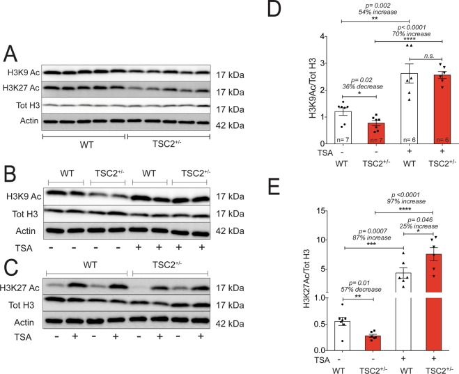
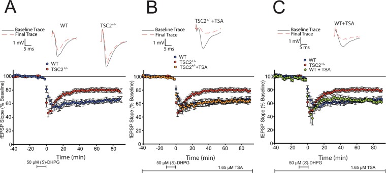
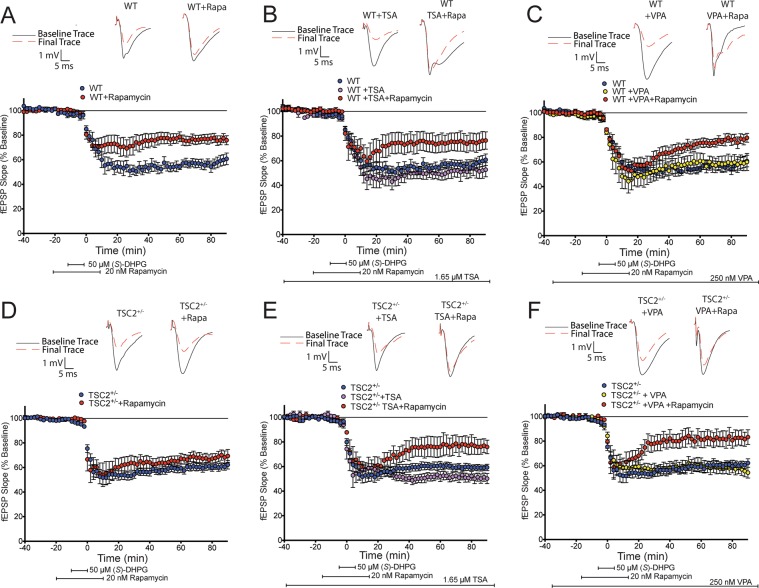
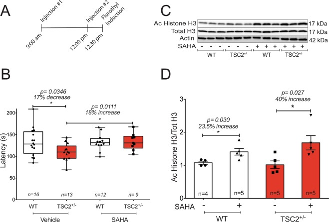
Abnormal synaptic plasticity has been implicated in several neurological disorders including epilepsy, dementia and Autism Spectrum Disorder (ASD). Tuberous Sclerosis Complex (TSC) is an autosomal dominant genetic disorder that manifests with seizures, autism, and cognitive deficits. The abnormal intracellular signaling underlying TSC has been the focus of many studies. However, nothing is known about the role of histone modifications in contributing to the neurological manifestations in TSC. Dynamic regulation of chromatin structure via post translational modification of histone tails has been implicated in learning, memory and synaptic plasticity. Histone acetylation and associated gene activation plays a key role in plasticity and so we asked whether histone acetylation might be dysregulated in TSC. In this study, we report a general reduction in hippocampal histone H3 acetylation levels in a mouse model of TSC2. Pharmacological inhibition of Histone Deacetylase (HDAC) activity restores histone H3 acetylation levels and ameliorates the aberrant plasticity in TSC2+/- mice. We describe a novel seizure phenotype in TSC2+/- mice that is also normalized with HDAC inhibitors (HDACis). The results from this study suggest an unanticipated role for chromatin modification in TSC and may inform novel therapeutic strategies for TSC patients.


Conflict of interest statement

The authors declare no competing interests.

Figures

Figure 1
Figure 1
TSC2+/− mouse hippocampi exhibit decreased global histone acetylation levels. (A) Representative cropped western blots of acute hippocampal slices acquired from adult WT and TSC2+/− mice (n = 5 animals per genotype). Each lane represents hippocampal lysate from a single animal. Hippocampal slices were harvested following incubation in artificial cerebrospinal fluid (ACSF) for 4 hours. Representative cropped western blots depicting H3K9 Ac protein levels (B) and H3K27 Ac (C) in acute hippocampal slices from adult WT and TSC2+/− animals harvested either in the presence or absence of the HDAC inhibitor, TSA (1.65 µM). Quantification of H3K9 Ac (D) and H3K27 Ac (E) protein levels from acutely harvested hippocampal slices from adult WT and TSC2+/− mice treated with or without TSA.
Figure 2
Figure 2
HDAC inhibition restores WT-like STP in response to 1 × TBS in adult TSC2+/− mice. A 1 × TBS elicits STP in adult WT hippocampal slices (shown in blue; n = 11 slices from 5 mice), while it provokes long lasting LTP in adult TSC2+/− mice (shown in red; n = 9 slices from 5 mice; two-way ANOVA: F(1,17) = 10.45, p = 0.0049). TSC2+/− slices incubated in TSA (1.65 µM) exhibited STP that was indistinguishable from untreated WT slices (n = 7 slices from 5 mice; two-way ANOVA: F(1,15) = 0.0007718, p = 0.9782). WT slices treated with TSA display long lasting LTP similar to that seen in TSC2+/− hippocampal slices (shown in purple; n = 6 slices from 6 mice; two-way ANOVA: F(1.13) = 1.557, p = 0.2350). TSA was introduced to the slices 60 minutes prior to 1 × TBS and was kept on for the duration of the experiment.
Figure 3
Figure 3
HDAC inhibition restores mGluR-LTD in juvenile TSC2+/− mice to mimic a juvenile WT response. (A) Acute hippocampal slices obtained from juvenile TSC2+/− mice (shown in red; n = 14 slices from 6 mice) exhibit a decreased mGluR-LTD magnitude compared to age matched littermate WT mice (shown in blue; n = 12 slices from 5 mice; two-way ANOVA: F(1,19) = 7.079, p < 0.0001). (B) Juvenile TSC2+/− slices treated with TSA (1.65 µM) display an mGluR-LTD that is indistinguishable from untreated juvenile WT slices (shown in orange; n = 8 slices from 4 mice; two-way ANOVA: F(1,13) = 0.7724, p = 0.7724). (C) Juvenile WT slices treated with TSA do not exhibit altered mGluR-LTD magnitude (shown in green; n = 5 slices from 5 mice); two-way ANOVA: F(1,10) = 0.02719, p = 0.8723).
Figure 4
Figure 4
HDAC inhibition restores mTORC1 dependent mGluR-LTD in adult TSC2+/− mice. (A) Adult WT mouse hippocampal slices (6–8 weeks old) exhibit LTD with (S)-DHPG (50 µM for 10 minutes; shown in blue, n = 5 slices from 4 mice) that is reduced in magnitude with rapamycin (20 nM for 20 minutes; shown in red, n = 8 slices from 4 mice; two-way ANOVA: F(1,15) = 7.375, p = 0.0159).The addition of the class I HDAC inhibitors, TSA (B, 1.65 µM; shown in purple, n = 5 slices from 4 mice) and VPA (C, 250 nM; shown in yellow, n = 6 slices from 4 mice) does not alter the acquisition of mGluR-LTD (B: two-way ANOVA: F(1.9) = 2.081, p = 0.1831. C: two-way ANOVA: F(1,11) = 0.1725, p = 0.6858) nor does it affect rapamycin sensitivity in adult WT hippocampal slices (shown in red for both; B: n = 7 slices from 4 mice; two-way ANOVA: F(1,11) 12.27, p = 0.0050; C: n = 7 slices from 5 mice; two-way ANOVA: F(1,10) = 9.629, p = 0.0112). D) Adult TSC2+/− mice exhibit LTD with (S)-DHPG (shown in blue, n = 8 slices from 4 mice) that is not sensitive to rapamycin (shown in red, n = 9 slices from 5 mice). E) Acquisition of mGluR-LTD is not altered in TSC2+/− slices treated with TSA (untreated slices shown in blue, n = 8 slices from 5 mice; TSA treated slices shown in purple n = 8 slices from 5 mice; two way ANOVA: F(1,18) = 1.271, p = 0.2743). TSC2+/− slices treated with TSA exhibit rapamycin sensitivity (shown in red, n = 6 slices from 5 mice; two-way ANOVA: F(1,12) = 7.566, p = 0.0176). F) VPA also restores rapamycin sensitivity in adult TSC2+/− slices (shown in red, n = 9 slices from 5 mice) and produces a decreased magnitude compared to slices treated with only VPA (shown in yellow, n = 7 slices from 5 mice; two-way ANOVA: F(1,14) = 6.054, p = 0.0275).
Figure 5
Figure 5
Juvenile TSC2+/− mice exhibit a reduced seizure threshold that is restored to WT-like latency with SAHA. (A) A schematic of the injection paradigm used in this study. Juvenile WT and TSC2+/− mice were intraperitoneally injected with either 50 mg/kg SAHA or vehicle (100 mM HPβCD) at the time of injection. Immediately following flurothyl induced seizures, a subset of mice was sacrificed, and whole hippocampal and cortical tissue was collected for post hoc western blot analysis. (B) Juvenile TSC2+/− mice exhibit a reduced seizure threshold in response to flurothyl compared to age matched WT mice (Student t-test: p = 0.0346). Compared to those treated with vehicle (HPβCD), SAHA increased latency to GTCS in juvenile TSC2+/− mice (Student t-test: p = 0.01) to levels that are indistinguishable from untreated WT mice. Compared to vehicle treated WT mice, WT mice treated with SAHA do not exhibit changes in latency to GTCS. (C) Following flurothyl induction, the hippocampi of vehicle or SAHA treated mice were rapidly harvested and processed for western blot analysis. Representative cropped western blot of whole hippocampal lysate extracted from vehicle or SAHA treated mice post flurothyl induction shows a global increase in acetylated histone H3 protein in SAHA treated mice, suggesting SAHA crossed the blood brain barrier and had a physiological effect. Each lane represents lysate from a single animal. An n = 3 per condition is represented in the blot. (D) Whole hippocampal extracts acquired from juvenile WT and TSC2+/− mice injected with either SAHA or vehicle were subject to western blot analysis. Quantification of protein levels shows that compared to vehicle treated mice, SAHA treatment increases pan acetyl histone H3 protein levels in the hippocampus of both WT mice (Student t-test:p = 0.03) and TSC2+/− mice (Student t-test: p = 0.027).

Comment in

References

    1. Takeuchi T, Duszkiewicz A, Morris R. The synaptic plasticity and memory hypothesis: encoding, storage and persistence. Philosophical Transactions of the Royal Society of London B: Biological Sciences. 2014;369:20130288. doi: 10.1098/rstb.2013.0288. - DOI - PMC - PubMed
    1. Martin, S., Grimwood, P. & Morris, R. Synaptic Plasticity and Memory: An Evaluation of the Hypothesis. Annual review of neuroscience 649–711, 10.1146/annurev.neuro.23.1.649 (2000). - PubMed
    1. Hebb, D. O. The organization of Behavior. John Wiley & Sons, Inc. (John Wiley & Sons, Inc., 1949).
    1. Bliss T, Collingridge G. A synaptic model of memory: long-term potentiation in the hippocampus. Nature. 1993;361:31–39. doi: 10.1038/361031a0. - DOI - PubMed
    1. Ebert D, Greenberg M. Activity-dependent neuronal signalling and autism spectrum disorder. Nature. 2013;493:327–337. doi: 10.1038/nature11860. - DOI - PMC - PubMed

Publication types

MeSH terms

Substances