. 2019 Mar 13:10:207.
doi: 10.3389/fneur.2019.00207. eCollection 2019.

Segment Occlusion vs. Reconstruction-A Single Center Experience With Endovascular Strategies for Ruptured Vertebrobasilar Dissecting Aneurysms

Affiliations

Segment Occlusion vs. Reconstruction-A Single Center Experience With Endovascular Strategies for Ruptured Vertebrobasilar Dissecting Aneurysms

Stefan Schob et al. Front Neurol. .

Abstract

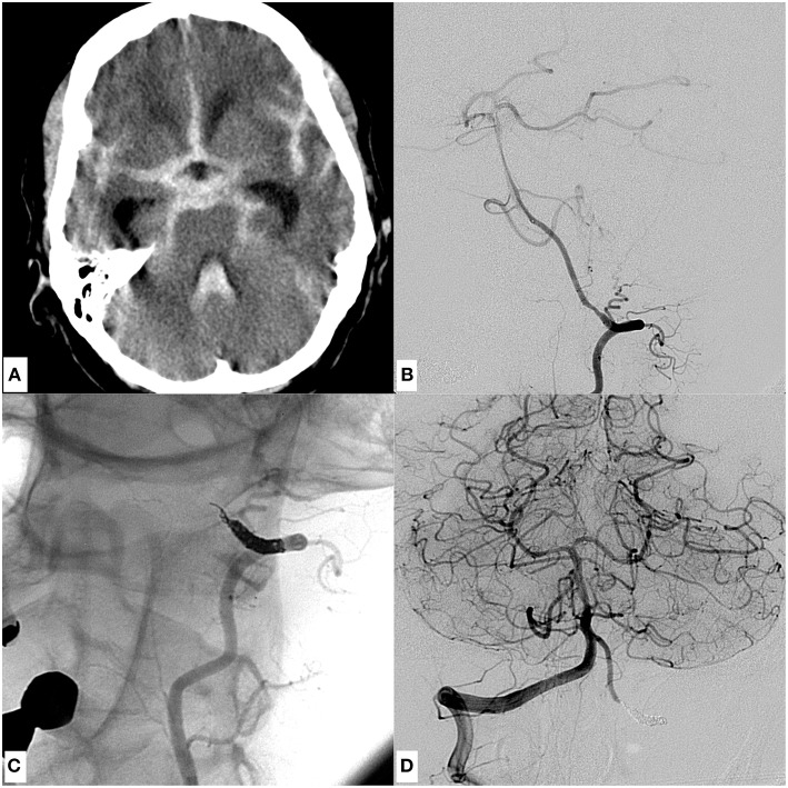
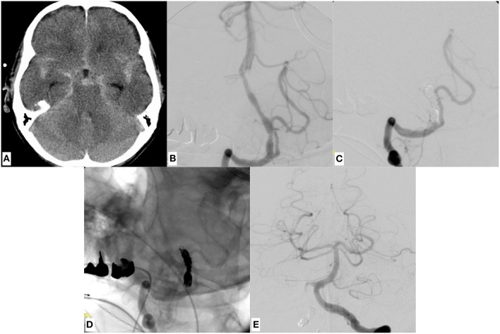
Objective: Ruptured dissecting aneurysms of the intracranial vertebral arteries exhibit an extraordinarily high risk for morbidity and mortality and are prone to re-rupture. Therefore, early treatment is mandatory to induce stagnation of the critical dynamic mural process. Appropriate endovascular approaches are segment sacrifice and reconstruction, however, both carry specific risks and benefits. To date most studies discuss only one of these approaches and focus on one specific device or technique. Therefore, our study aimed to present our experiences with both techniques, providing a considered approach on when to perform endovascular reconstruction or sacrifice. Materials and Methods: We retrospectively reviewed patients with subarachnoid hemorrhage in our database, suffering from dissecting aneurysms of the intradural vertebral arteries and treated endovascularly in the acute setting. A total of 16 cases were included. Clinical history, radiologic findings and outcomes were analyzed. Results: In 7 patients a reconstructive approach was chosen with 4 of them receiving stent-assisted coiling as primary strategy. One of the 7 patients suffered early re-bleeding due to progression of the dissection and therefore treatment was augmented with implantation of 2 flow diverters. The remaining 2 patients were primarily treated with flow diverters in telescoping technique. In 9 patients a deconstructive approach was followed: 6 patients were treated with proximal coil-occlusion of the V4 segment, 3 patients received distal coiling of the V4 segment. Two patients died (GOS 1) in the subacute stage due to sequelae of recurrent episodes of raised intracranial pressure and parenchymal hemorrhage. Two patients kept severe disability (GOS 3), six patients had moderate disability (GOS 4) and seven patients showed full recovery (GOS 5). None of the patients suffered from a procedural or postprocedural ischemic stroke. Conclusions: In patients with good collateral vascularization, proximal, or distal partial segment sacrifice via with endovascular coil occlusion seems to yield the best risk-benefit ratio for treatment of ruptured dissecting V4 aneurysms, especially since no continued anticoagulation is required and possibly essential surgery remains feasible in this scenario. If possible, PICA occlusion should be avoided-although even proximal PICA occlusion can become necessary, when weighing against the risk of an otherwise untreated ruptured V4 dissecting aneurysm. Contrarily, if the dominant V4 segment is affected, the hemodynamic asymmetry prohibits occlusion and necessitates reconstruction of the respective segment. For this, implants with high metal coverage treating the entire affected segment appear to be the most promising approach.

Keywords: dissecting aneurysm; flow diverter; segment occlusion; stent assisted coiling; subarachnoid hemorrhage; vertebral artery.


Figures

Figure 1
Figure 1
An example of a dissecting V4 aneurysm causative for SAH Fisher grade 4, Hunt and Hess grade 4, treated with proximal endovascular segment occlusion. (A) Shows a representative slice of the initial CCT with significant SAH. (B) Demonstrates the mural injury of the hypoplastic left-sided intradural vertebral artery, visible as tapered narrowing of the proximal V4 segment in an oblique projection of a left vertebral artery injection. (C) Image after proximal segment occlusion in magnified working projection: two coil loops of the first coil were anchored intraluminally behind the distal end of the dissected V4 segment in corkscrew technique, preventing retrograde reperfusion of the dissection site. Note the elliptically shaped, condensed proximal coil bundle which occludes the proximal starting point of the dissection, solidly impeding its anterograde reperfusion. (D) Depicts the right vertebral artery angiogram, showing distinct retrograde perfusion of the remaining left V4 segment, thus securing sufficient supply of small brain stem perforators. Symmetric contrast filling of both cerebellar hemispheres directly after coiling, indicating sufficient blood flow into the entire posterior circulation through the unaffected right vertebral artery. The patient did not suffer rebleeding and showed excellent recovery 3 months after the event (GOS 5).
Figure 2
Figure 2
A representative case of a dissecting V4 aneurysm (SAH Fisher grade 4, Hunt and Hess grade 4) of the right sided, small-sized vertebral artery, treated with distal endovascular segment occlusion. (A) Shows the initial CCT revealing SAH. (B) Depicts the right vertebral artery pathology, visible as pearl and string sign in DSA. Note the significant PICA branching proximal to the affected V4 portion. (C,D) Provide substracted and unsubstracted images of right vertebral artery injections after distal segment occlusion, leaving the PICA orifice open. Strong opacification of the now functionally PICA-terminating V4 segment. The first coil, which was anchored in corkscrew technique within the true lumen of the distal V4, is securing the dissecting aneurysm whilst impeding anterograde or retrograde reperfusion. (E) The contralateral vertebral artery angiogram, showing symmetrical opacification of the entire posterior territory after occlusion of the right-sided dissected V4 segment. The patient did not suffer rebleeding and showed excellent recovery 2 months after the event (GOS 5).
Figure 3
Figure 3
An example of SAH due to a ruptured dissecting aneurysm of the right sided, hemodynamically dominant vertebral artery involving the PICA orifice. It was finally managed through stent assisted coiling using a laser cut device (Enterprise stent, Codman Neuro, USA). (A) Demonstrates basal SAH. (B) Shows a DSA image of a right vertebral artery injection revealing the causative large dissecting aneurysm, which was treated with Enterprise stent—assisted coiling (D) in jailing technique. (C) Radiopaque markers of the enterprise stent proximal and distal to the aneurysm. (D,E) Show the successful reconstruction of the affected V4 segment with continuous opacification of the PICA. Note the placement of the micro-guide-wire in the proximal PICA. (C–E) The patient regained self-reliance in the daily routine (GOS 4) 4 months after the event.
Figure 4
Figure 4
A case of SAH due to a ruptured dissecting aneurysm of the dominant left sided vertebral artery of a 53 years old male patient, which was initially treated with stent assisted coiling using a braided stent (LEO, Balt, France). (A) Provides a representative CCT section showing SAH Fisher grade 4. (B,C) Give the anteroposterior and lateral DSA images revealing the underlying, morphologically subtle dissecting aneurysm close to the PICA orifice. The inferior row of images (D–F) shows the LEO stent and the adjacent coil bundle after treatment in situ without (D) and with injection of contrast agent (E), demonstrating unremarkable vertebrobasilar opacification (F) (see Figure 5).
Figure 5
Figure 5
The complicated further course of the patient initially treated with stent assisted coiling (Figure 4). 7 days after the first procedure the patient complained of sudden, predominantly left sided, most severe head and neck pain. Therefore, a CCT was performed (A) and showed a significant subarachnoid rebleeding predominantly in the basal cisterns. Note the clearly depictable radiopaque LEO stent in the left V4 segment and the surrounding hyperdense subarachnoid blood. Images (B,C) show the morphologically subtle progress of the previously treated dissecting aneurysm of the left V4 segment. To secure the further distended dissecting aneurysm—whose proximal and distal endings were not precisely definable—two overlapping flow diverters (p64, Phenox, Germany) with highly increased surface coverage in comparison to the priorly implanted LEO stent were used to safely overlay the whole affected segment (D) in telescoping technique. Note the eight circular radiopaque markers at the proximal ending of both flow diverters in (D), which are now spanning the proximal and distal end of the previously implanted LEO stent. Images (E,F) show the successfully reconstructed vertebral artery with delayed but complete filling of the ipsilateral PICA. The patient did not suffer further rebleeding and regained self-reliance in the daily routine (GOS 4) 2 months after the event.
Figure 6
Figure 6
A patient being treated with 3 flow diverters plus coiling in jailing technique (Fisher 4, Hunt and Hess 4). CCT (A) had shown a significant subarachnoid hemorrhage with accentuation in the posterior fossa. Image (B) shows the underlying dissecting aneurysm of the left sided, dominant vertebral artery located in the terminal V4 segment. (C) PICA-terminating contralateral vertebral artery, without sufficient collateral circulation to the posterior circulation. To promote coagulation within the pseudoaneurysm, 2 coils were placed carefully within the aneurysm sac after implantation of 3 overlapping PED 2 Flex flow diverters (D,E). The dissecting aneurysm was highly fragile as indicated by the subtle growth of the false lumen during the intervention (most obvious when comparing the morphology of the pseudoaneurysm in (B,E). (F) Provides the final injection after successful reconstruction of the segment. The patient did not suffer further rebleeding and is recovering from the hemorrhagic event (GOS 4).
Figure 7
Figure 7
Interventional algorithm for endovascular treatment of dissecting V4 aneurysms especially considering the hemodynamic situation at hand. Green panels represent the most favorable option. Arrow thickness indicates higher preferability of the respective approach.

References

    1. Debette S, Compter A, Labeyrie MA, Uyttenboogaart M, Metso TM, Majersik JJ, et al. . Epidemiology, pathophysiology, diagnosis, and management of intracranial artery dissection. Lancet Neurol. (2015) 14:640–54. 10.1016/S1474-4422(15)00009-5 - DOI - PubMed
    1. Yamada M, Miyasaka Y, Yagishita S, Fujii K. Dissecting aneurysm of the intracranial vertebral artery associated with proximal focal degeneration of the elastica: a comparative pathological study of the vertebral artery in patients with and without aneurysms. Surg Neurol. (2003) 60:431–7; discussion 437. 10.1016/S0090-3019(03)00324-0 - DOI - PubMed
    1. Hong JM, Chung CS, Bang OY, Yong SW, Joo IS, Huh K. Vertebral artery dominance contributes to basilar artery curvature and peri-vertebrobasilar junctional infarcts. J Neurol Neurosurg Psychiatry. (2009) 80:1087–92. 10.1136/jnnp.2008.169805 - DOI - PMC - PubMed
    1. Boström K, Liliequist B. Primary dissecting aneurysm of the extracranial part of the internal carotid and vertebral arteries. A report of three cases. Neurology. (1967) 17:179–86. - PubMed
    1. Arnold M, Bousser MG. Clinical manifestations of vertebral artery dissection. Front Neurol Neurosci. (2005) 20:77–86. 10.1159/000088152 - DOI - PubMed

LinkOut - more resources