Uterine arteriovenous malformation with repeated vaginal bleeding after dilatation and curettage
- PMID: 30918884
- PMCID: PMC6422849
- DOI: 10.5468/ogs.2019.62.2.142
Uterine arteriovenous malformation with repeated vaginal bleeding after dilatation and curettage
Abstract
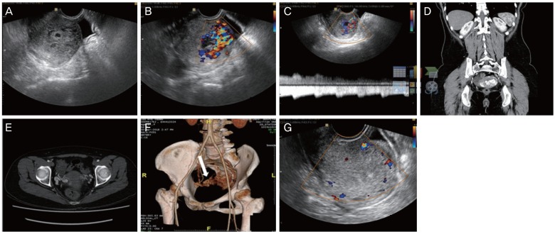
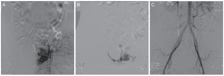
Uterine arteriovenous vascular malformation (UAVM) is a disease that causes excessive bleeding. The symptoms do not subside without proper treatment and this can lead to life-threatening situations. The correct diagnosis of UAVM can be complicated if the patient's uterus did not completely discharge everything during abortion (in broader terms, retaining remnants of the products of conception). In this case, Doppler ultrasonography and computed tomography angiography with 3-dimensional rendering were used to analyze the cause of bleeding and provide proper treatment of this patient. Then, uterine artery embolization, dilatation, and curettage were performed safely and successfully. The patient no longer had symptoms of vaginal spotting during the planned follow up care. UAVM is uncommon; however, if reproductive-age women show repeated abnormal vaginal bleeding after dilatation and curettage, a diagnosis of UAVM must be considered based on the medical history and examination.
Keywords: Arteriovenous malformation; Ultrasonography; Uterine artery; Vaginal bleeding.
Conflict of interest statement
Conflict of interest: No potential conflict of interest relevant to this article was reported.
Figures
Similar articles
-
Ultrasound diagnosis and management of acquired uterine enhanced myometrial vascularity/arteriovenous malformations.Am J Obstet Gynecol. 2016 Jun;214(6):731.e1-731.e10. doi: 10.1016/j.ajog.2015.12.024. Epub 2016 Feb 9. Am J Obstet Gynecol. 2016. PMID: 26873276
-
Acquired uterine arteriovenous malformation following dilation and curettage: a case report.Pan Afr Med J. 2022 May 26;42:71. doi: 10.11604/pamj.2022.42.71.35371. eCollection 2022. Pan Afr Med J. 2022. PMID: 36034014 Free PMC article.
-
A case report of Ggeneralized uterine arteriovenous malformation after molar pregnancy in an infertile woman.Int J Reprod Biomed. 2018 Feb;16(2):119-122. Int J Reprod Biomed. 2018. PMID: 29675497 Free PMC article.
-
Uterine arteriovenous malformation (UAVM) as a rare cause of postpartum hemorrhage (PPH): a literature review.Arch Gynecol Obstet. 2022 Dec;306(6):1873-1884. doi: 10.1007/s00404-022-06498-0. Epub 2022 Mar 13. Arch Gynecol Obstet. 2022. PMID: 35284958 Review.
-
Uterine arteriovenous malformation - diagnosis and management.Ginekol Pol. 2018;89(5):276-279. doi: 10.5603/GP.a2018.0047. Ginekol Pol. 2018. PMID: 30084480 Review.
Cited by
-
Sonographic Features of Uterine Arteriovenous Malformation: A Case Series.Diagnostics (Basel). 2024 Apr 23;14(9):873. doi: 10.3390/diagnostics14090873. Diagnostics (Basel). 2024. PMID: 38732288 Free PMC article.
-
Embolization of uterine arteriovenous malformation causing postpartum hemorrhage using n-butyl cyanoacrylate: A case report.Radiol Case Rep. 2021 Mar 19;16(5):1188-1190. doi: 10.1016/j.radcr.2021.02.053. eCollection 2021 May. Radiol Case Rep. 2021. PMID: 33777284 Free PMC article.
-
Uterine Arteriovenous Malformation: Diagnostic and Therapeutic Challenges.Diagnostics (Basel). 2024 May 23;14(11):1084. doi: 10.3390/diagnostics14111084. Diagnostics (Basel). 2024. PMID: 38893611 Free PMC article.
References
-
- Grivell RM, Reid KM, Mellor A. Uterine arteriovenous malformations: a review of the current literature. Obstet Gynecol Surv. 2005;60:761–767. - PubMed
-
- Ahn HY, Park IY, Lee G, Kim SJ, Shin JC. Uterine arteriovenous malformation. Arch Gynecol Obstet. 2005;271:172–175. - PubMed
-
- Oguz Y, Gonca Eldem F, Cil B, Sanhal C, Gençosmanoğlu-Türkmen G, Aykan Y, et al. Uterine arteriovenous malformation. Gynecol Obstet Reprod Med. 2018
-
- Polat P, Suma S, Kantarcý M, Alper F, Levent A. Color Doppler US in the evaluation of uterine vascular abnormalities. Radiographics. 2002;22:47–53. - PubMed
Publication types
LinkOut - more resources
Full Text Sources