. 2019 Jul;95(7):722-725.
doi: 10.1002/cyto.a.23753. Epub 2019 Mar 28.

OMIP-056: Evaluation of Human Conventional T Cells, Donor-Unrestricted T Cells, and NK Cells Including Memory Phenotype by Intracellular Cytokine Staining

Affiliations

OMIP-056: Evaluation of Human Conventional T Cells, Donor-Unrestricted T Cells, and NK Cells Including Memory Phenotype by Intracellular Cytokine Staining

One Dintwe et al. Cytometry A. 2019 Jul.

Erratum in

Abstract

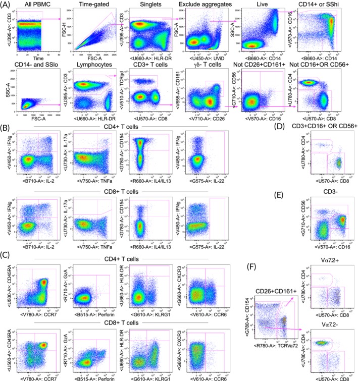
A 26-color staining panel was developed to profile human antigen-specific T cells in an intracellular cytokine staining (ICS) assay using peptide pools to various antigens of interest. In addition to multiple functional markers, the panel includes differentiation/activation markers and markers to assess γδ, mucosal-associated invariant T, and NK T cells as well as conventional NK cells. Panel optimization was performed using previously cryopreserved PBMC from healthy adults, and then, expression of key functional markers in the panel was cross-validated against a validated ICS assay used in the HIV Vaccine Trials Network (HVTN). The panel is currently being used to evaluate the responses to tuberculosis and malaria vaccine candidates in volunteers from different geographic areas. © 2019 The Authors. Cytometry Part A published by Wiley Periodicals, Inc. on behalf of International Society for Advancement of Cytometry.

Keywords: MAIT cells; NK cells; T cells; cytometry; human PBMC; intracellular cytokines; memory; γδ T cells.


Figures

Figure 1
Figure 1
Example of the staining and gating strategy for PBMC stimulated with staphylococcal enterotoxin B (SEB). PBMC from a healthy adult were stimulated for 6 h with SEB. (A) Gating hierarchy to identify lineages. Initial gating on time (seconds) to exclude any events early in collection if there are pressure fluctuations, singlet gating on forward scatter height vs. area, exclusion of aggregates (only one example shown, but several sequential gates on various parameters are used), and live cell gating. Monocytes are gated as either CD14+ or high for side scatter and the upper right graph shows three monocyte subsets based on CD14 vs. CD16. Non‐monocytes are gated as CD14‐SSlo and then scatter gated on lymphocytes. The gating scheme avoids any overlapping subsets as shown in supplemental figure 4. Thus, conventional CD4+ and CD8+ T cells are gated as CD3+, then γδ‐, not CD26 + CD161+ (containing MAIT cells), and not CD16+ OR CD56+ (containing NKT cells). (B) Functional markers for CD4+ and CD8+ T cells. A gate is applied for each cytokine, and Boolean gates are created to identify cells expressing different combinations of markers. The single function gates are sometimes chosen vs. a parameter that displays some FMO spreading to allow for angled gates. Most gates are copied, applied to all lineages, and then cloned so that any changes to the gate on one lineage changes that gate for all lineages. However, the IL‐22 gate was uniquely lower for CD4 T cells (compared to other lineages) since the CD8 reagent caused some spreading into IL‐22 and thus requiring a higher gate for all other lineages that express CD8. (C) Additional functional and non‐functional markers for CD4+ and CD8+ T cells. Perforin and granzyme A are constitutive but can be examined as co‐expression with another functional marker. (D) NK T cells gated as CD16+ OR CD56+ on CD3+ γδ‐ T cells. Expression of CD4 vs. CD8 is shown, but all the other markers are also gated on the NK T cells. (E) NK cell subsets defined by CD16 vs. CD56 on CD3‐ lymphocytes. (F) MAIT cells identified as CD3+ γδ‐ CD26 + CD161+ and then Vα7.2+. The Va7.2+ cells are predominantly CD8+; however, the Vα7.2‐ cells are predominantly CD4+ and are likely not MAIT cells. For all gates, none are placed lower than that defined by FMO controls. Some gates are placed higher to improve the specificity, for example, for the functional markers based on the background as observed in the unstimulated controls (Online Fig. 6). The labels above each graph indicate the cells included in that graph.

References

    1. De Rosa SC, Carter DK, McElrath MJ. OMIP‐014: Validated multifunctional characterization of antigen‐specific human T cells by intracellular cytokine staining. Cytometry A 2012;81:1019–1021. - PMC - PubMed
    1. Moncunill G, Dobano C, McElrath MJ, De Rosa SC. OMIP‐025: Evaluation of human T‐ and NK‐cell responses including memory and follicular helper phenotype by intracellular cytokine staining. Cytometry A 2015;87:289–292. - PMC - PubMed

Publication types

MeSH terms

LinkOut - more resources