

Extracutaneous manifestations in phacomatosis cesioflammea and cesiomarmorata: Case series and literature review
- PMID: 30920161
- PMCID: PMC6488410
- DOI: 10.1002/ajmg.a.61134
Extracutaneous manifestations in phacomatosis cesioflammea and cesiomarmorata: Case series and literature review
Abstract
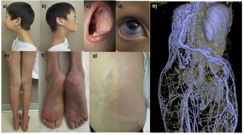
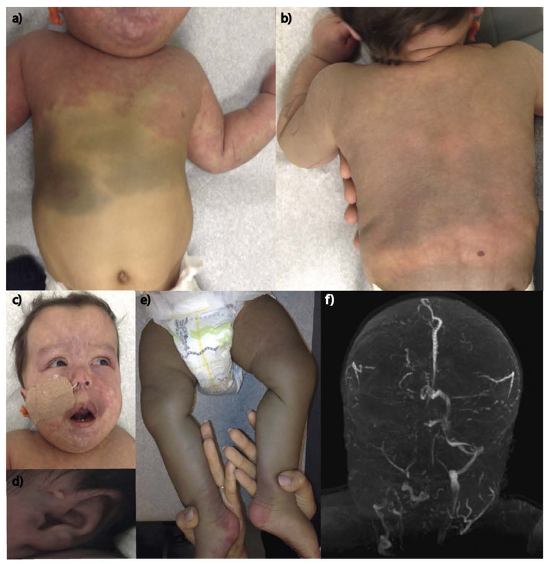
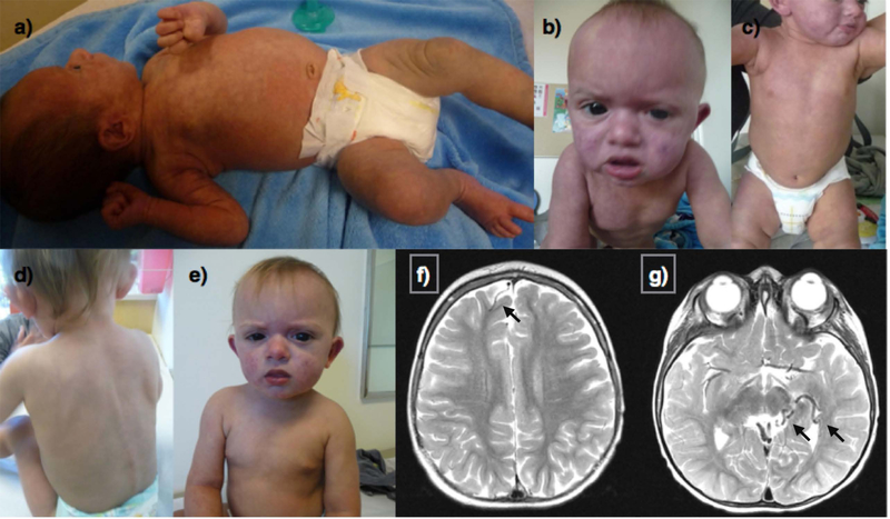
Phacomatosis pigmentovascularis (PPV) comprises a family of rare conditions that feature vascular abnormalities and melanocytic lesions that can be solely cutaneous or multisystem in nature. Recently published work has demonstrated that both vascular and melanocytic abnormalities in PPV of the cesioflammea and cesiomarmorata subtypes can result from identical somatic mosaic activating mutations in the genes GNAQ and GNA11. Here, we present three new cases of PPV with features of the cesioflammea and/or cesiomarmorata subtypes and mosaic mutations in GNAQ or GNA11. To better understand the risk of potentially occult complications faced by such patients we additionally reviewed 176 cases published in the literature. We report the frequency of clinical findings, their patterns of co-occurrence as well as published recommendations for surveillance after diagnosis. Features assessed include: capillary malformation; dermal and ocular melanocytosis; glaucoma; limb asymmetry; venous malformations; and central nervous system (CNS) anomalies, such as ventriculomegaly and calcifications. We found that ocular findings are common in patients with phacomatosis cesioflammea and cesiomarmorata. Facial vascular involvement correlates with a higher risk of seizures (p = .0066). Our genetic results confirm the role of mosaic somatic mutations in GNAQ and GNA11 in phacomatosis cesioflammea and cesiomarmorata. Their clinical and molecular findings place these conditions on a clinical spectrum encompassing other GNAQ and GNA11 related disorders and inform recommendations for their management.
Keywords: GNA11; GNAQ; management; phacomatosis cesioflammea; phacomatosis cesiomarmorata; phacomatosis pigmentovascularis.
© 2019 Wiley Periodicals, Inc.
Figures
References
Publication types
MeSH terms
Substances
Supplementary concepts
Grants and funding
- U01 HG007943/NH/NIH HHS/United States
- U01 HG007690/HG/NHGRI NIH HHS/United States
- U01 TR001395/NH/NIH HHS/United States
- U01 HG007708/HG/NHGRI NIH HHS/United States
- U01 HG007672/HG/NHGRI NIH HHS/United States
- U01 HG007690/NH/NIH HHS/United States
- U54 NS093793/NH/NIH HHS/United States
- U01 HG007943/HG/NHGRI NIH HHS/United States
- U01 HG007942/HG/NHGRI NIH HHS/United States
- U01 HG007674/NH/NIH HHS/United States
- U01 HG007674/HG/NHGRI NIH HHS/United States
- U01 HG007709/NH/NIH HHS/United States
- U01 HG007703/NH/NIH HHS/United States
- U01 HG007672/NH/NIH HHS/United States
- U01 HG010218/HG/NHGRI NIH HHS/United States
- U01 TR001395/TR/NCATS NIH HHS/United States
- U01 HG007709/HG/NHGRI NIH HHS/United States
- U54 NS093793/NS/NINDS NIH HHS/United States
- U01 HG007530/HG/NHGRI NIH HHS/United States
- U01 HG007703/HG/NHGRI NIH HHS/United States
- U01 HG007530/NH/NIH HHS/United States
