Review
. 2019 Aug;39(8):459-471.
doi: 10.1089/jir.2019.0014. Epub 2019 Mar 28.

Inflammation and Immunity: From an Adipocyte's Perspective

Affiliations
Review

Inflammation and Immunity: From an Adipocyte's Perspective

Calvin C Chan et al. J Interferon Cytokine Res. 2019 Aug.

Abstract

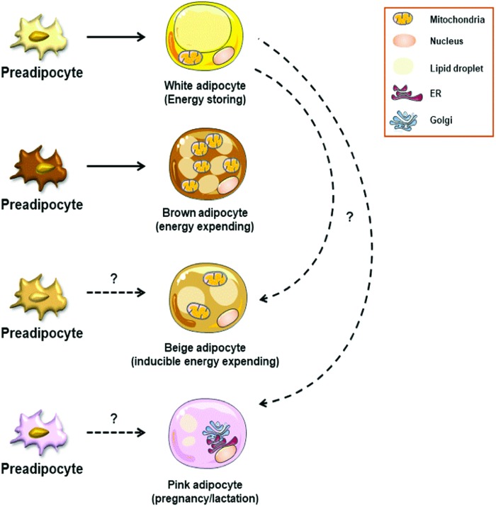
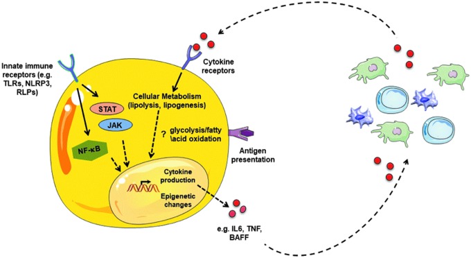
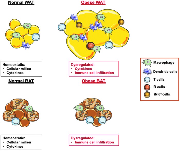
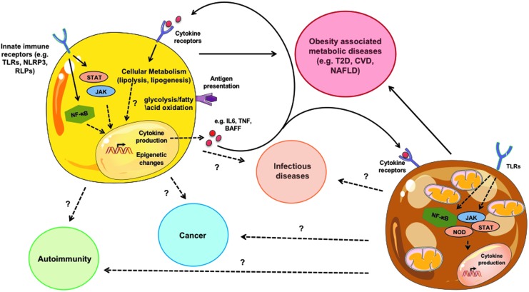
Comprehension of adipocyte function has evolved beyond a long-held belief of their inert nature, as simple energy storing and releasing cells. Adipocytes, including white, brown, and beige, are capable mediators of global metabolic health, but their intersection with inflammation is a budding field of exploration. Evidence hints at a reciprocal relationship adipocytes share with immune cells. Adipocyte's capacity to behave in an "immune-like" manner and ability to sense inflammatory cues that subsequently alter core adipocyte function might play an important role in shaping immune responses. Clarifying this intricate relationship could uncover previously underappreciated contribution of adipocytes to inflammation-driven human health and disease. In this review, we highlight the potential of largely underappreciated adipocyte "immune-like" function and how it may contribute to inflammation, immunity, and pathology of various diseases.

Keywords: adipocytes; cytokines; inflammation; interferon; metabolism; obesity.


Conflict of interest statement

The authors declare no competing financial interests.

Figures

<b>FIG. 1.</b>
FIG. 1.
Adipocyte subsets. Adipocytes are divided into various subtypes: white adipocytes (energy storing), brown adipocytes (thermogenesis), beige/brite adipocytes (inducible energy expending), and pink adipocytes (pregnancy/lactation associated). White adipocytes are characterized by large lipid droplets and are known for their energy storing capacity during obesity. Brown adipocytes are characterized by numerous mitochondria. They originate from a separate preadipocyte population. Mitochondrial number and function within brown adipocytes support their thermogenic capacity and maintenance of body temperature. Beige adipocytes arise within WAT and are characterized by unique thermogenic capacity. Induction of beige adipocytes is driven by environmental cues (eg, cold stress, exercise, and β3-adrenergic stimulation). The developmental origins of beige adipocytes are still under investigation. Pink adipocytes are proposed to transdifferentiate from cells within subcutaneous AT. The precise function and developmental origins of pink adipocytes remains underdefined. WAT, white adipose tissue. Color images are available online.
<b>FIG. 2.</b>
FIG. 2.
Adipocyte-centric inflammation and crosstalk with immune cells. Adipocytes possess an “immune-like” potential, including (a) expression of innate immune receptors (eg, TLRs, NLRs, and RLRs), (b) cytokine (eg, TNF, IL1β, IL-6, BAFF, and APRIL) and chemokine (CCL2, CXCL8, CXCL1, CXCL10) production, and (c) antigen presentation (eg, MHC-II, CD80, CD40, CD1d, and CIITA). Adipocyte sensing of cytokines alter their function (eg, lipolysis, lipogenesis, and UCP-1 thermogenesis). Cumulatively, these adipocyte inflammatory features suggest potential crosstalk with immune cells (in AT or circulation) that may directly impact adipocyte function, immune cell function, and/or disease pathogenesis. TLR, toll-like receptor; NLR, NOD-like receptor; RLR, RIG-I receptor; BAFF, B cell activating factor; APRIL, a proliferation-inducing ligand; MHC, major histocompatibility complex. Color images are available online.
<b>FIG. 3.</b>
FIG. 3.
Obesity-driven AT inflammatory plasticity. White and brown AT in a lean state consists of a diverse cellular milieu (eg, adipocytes, macrophages, T cells, and B cells) and capacity to produce cytokines. Pathological expansion of AT in obesity promotes dysregulation of cytokine production (e.g., TNF, IL-6, IL-1, and IFN-γ), enhances immune cell infiltration/polarization/activation and exacerbates the AT inflammatory milieu. Although contribution of AT to obesity-associated inflammation is well investigated, adipocyte-specific contribution to inflammation is understudied and remains a critical gap in knowledge. Color images are available online.
<b>FIG. 4.</b>
FIG. 4.
Adipocyte contribution to disease. Adipocytes have “immune-like” capabilities that, in part, overlap with traditionally investigated immune cells. White adipocyte-specific inflammatory capacity can impact obesity-associated metabolic diseases. The contributions of other subsets of adipocytes (eg, brown) to disease should be formally examined. Relevance of the interconnection in adipocytes between signaling pathways (eg, NF-κB and Jak/STAT), core metabolism (e.g., glycolysis and β-oxidation), and epigenetic programming to promote adipocyte-intrinsic inflammatory capacity and contribute to disease pathogenesis warrants future investigation. In addition, if adipocyte crosstalk with immune cells likewise contributes to diseases, including autoimmunity, cancer and infections are a natural extension to current investigations. Color images are available online.

References

    1. Alexaki VI, Notas G, Pelekanou V, Kampa M, Valkanou M, Theodoropoulos P, Stathopoulos EN, Tsapis A, Castanas E. 2009. Adipocytes as immune cells: differential expression of TWEAK, BAFF, and APRIL and their receptors (Fn14, BAFF-R, TACI, and BCMA) at different stages of normal and pathological adipose tissue development. J Immunol 183(9):5948–5956 - PubMed
    1. Babaei R, Schuster M, Meln I, Lerch S, Ghandour RA, Pisani DF, Bayindir-Buchhalter I, Marx J, Wu S, Schoiswohl G, Billeter AT, Krunic D, Mauer J, Lee YH, Granneman JG, Fischer L, Muller-Stich BP, Amri EZ, Kershaw EE, Heikenwalder M, Herzig S, Vegiopoulos A. 2018. Jak-TGFbeta cross-talk links transient adipose tissue inflammation to beige adipogenesis. Sci Signal 11(527): pii: - PubMed
    1. Bae J, Ricciardi CJ, Esposito D, Komarnytsky S, Hu P, Curry BJ, Brown PL, Gao Z, Biggerstaff JP, Chen J, Zhao L. 2014. Activation of pattern recognition receptors in brown adipocytes induces inflammation and suppresses uncoupling protein 1 expression and mitochondrial respiration. Am J Physiol Cell Physiol 306(10):C918–C930 - PubMed
    1. Balaban S, Lee LS, Schreuder M, Hoy AJ. 2015. Obesity and cancer progression: is there a role of fatty acid metabolism? Biomed Res Int 2015:274585. - PMC - PubMed
    1. Ballak DB, van Asseldonk EJ, van Diepen JA, Jansen H, Hijmans A, Joosten LA, Tack CJ, Netea MG, Stienstra R. 2015. TLR-3 is present in human adipocytes, but its signalling is not required for obesity-induced inflammation in adipose tissue in vivo. PLoS One 10(4):e0123152. - PMC - PubMed

Publication types

LinkOut - more resources