Review
. 2019 Mar 29;124(7):1025-1044.
doi: 10.1161/CIRCRESAHA.118.313260.

Neurovascular and Cognitive Dysfunction in Hypertension

Affiliations
Review

Neurovascular and Cognitive Dysfunction in Hypertension

Costantino Iadecola et al. Circ Res. .

Abstract

Hypertension has emerged as a leading cause of age-related cognitive impairment. Long known to be associated with dementia caused by vascular factors, hypertension has more recently been linked also to Alzheimer disease-the major cause of dementia in older people. Thus, although midlife hypertension is a risk factor for late-life dementia, hypertension may also promote the neurodegenerative pathology underlying Alzheimer disease. The mechanistic bases of these harmful effects remain to be established. Hypertension is well known to alter in the structure and function of cerebral blood vessels, but how these cerebrovascular effects lead to cognitive impairment and promote Alzheimer disease pathology is not well understood. Furthermore, critical questions also concern whether treatment of hypertension prevents cognitive impairment, the blood pressure threshold for treatment, and the antihypertensive agents to be used. Recent advances in neurovascular biology, epidemiology, brain imaging, and biomarker development have started to provide new insights into these critical issues. In this review, we will examine the progress made to date, and, after a critical evaluation of the evidence, we will highlight questions still outstanding and seek to provide a path forward for future studies.

Keywords: Alzheimer’s disease; biomarkers; brain blood supply; dementia; risk factors; white matter pathology.


Figures

Figure 1:
Figure 1:
Cerebrovascular anatomy and segmental pathology induced by hypertension. Large extracranial cerebral arteries (internal carotid arteries and vertebral arteries) enter the skull and converge at the base of the brain to form the circle of Willis from which the major intracranial cerebral arteries emanate. Branches of these arteries form a dense anastomotic network (pial arteries ad arterioles) which give rise to arterioles that dive into the substance of the brain (penetrating arterioles). Some vessels, like the lenticulostriate arteries, arise from the circle of Willis and proximal branches and vascularize the deep brain structures including the white matter. The predominant pathology caused by hypertension in the different segment of the cerebrovascular tree is indicated. Due to its precarious blood supply from terminal arterioles particularly vulnerable to hypertensive damage, the subcortical white matter is highly likely to be harmed by hypoxia-ischemia. Abbreviations: ICA, internal carotid artery; MCA, middle cerebral artery; WM, white matter.
Figure 2:
Figure 2:
Cerebrovascular autoregulation and hypertension. Autoregulation keeps CBF relatively stable during fluctuations in BP. Autoregulation was first examined by measuring CBF during stepwise changes in BP, once a steady state is reached (static autoregulation) (left). Based largely on animal data, chronic hypertension was found to shift the pressure-flow curve to the right, such that the autoregulated range moves towards higher BP. The development of methods to assess CBF dynamically like transcranial doppler flowmetry, enabled to study the flow velocity changes in large cerebral arteries during BP transients induced by different maneuvers, e.g., standing from a sitting position. As illustrated in the right panel, MCA flow drops in sync with AP. But, owing to an autoregulatory drop in cerebrovascular resistance (CVR), it recovers faster. Evidence suggests that hypertension does not impair dynamic autoregulation, except in severe hypertension (see text for further details). Right panel data from Aaslid et al.
Figure 3:
Figure 3:
Putative mechanisms of neurovascular dysfunction in slow pressor AngII hypertension, BPH mice and DOCA-salt hypertension. Circulating or brain AngII reaches AT1R in PVM wherein it activates Nox2 leading the vascular oxidative stress and neurovascular dysfunction. AT1R and Nox2 are also present in other vascular cells, but they do not seem to play a primary role in these hypertension models. The cartoon depicts a penetrating arteriole, but brain macrophages are also present in the meninges, wherein they may play a role in pial and meningeal vessel vasomotor function and permeability. Whether PVM have a similar role in models of AngII-independent hypertension remains to be established. Abbreviations: AT1R, AngII type 1 receptor; PVM, perivascular macrophages; SMC, smooth muscle cells. (Illustration Credit: Ben Smith).
Figure 4:
Figure 4:
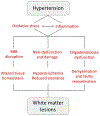
Potential mechanisms of WM damage by hypertension. Vascular oxidative stress and inflammation disrupt the BBB, induce NVU dysfunction and damage, and impair oligodendrocyte development and function. The resulting alterations in tissue homeostasis, hypoxia-ischemia, reduced brain clearance and impaired remyelination lead to WM damage. Abbreviations: NVU, neurovascular unit.
Figure 5:
Figure 5:
Brain lesions produced by hypertension that underlie cognitive impairment. Brain atrophy, microinfarcts and microbleeds cause neuronal loss and brain dysfunction. In addition, microinfarcts and microbleeds disrupt brain connectivity and reduce network efficiency Damage to WM tracts (WML) also degrades connectivity, especially in thalamo-cortico networks. Alteration in PVS impair brain clearance and may promote protein accumulation in brain and vessels, and dysfunction of the NVU leads to vascular insufficiency and BBB damage. Potential interactions between different pathogenic mechanisms are indicated by the dashed arrows. Abbreviations: NVU, neurovascular unit; PVS, perivascular spaces; WML, white matter lesions.
Figure 6:
Figure 6:
Potential mechanisms underlying the relationship between hypertension and AD. The vascular damage produced by hypertension leads to brain dysfunction thought hypoxia-ischemia, as well as increase in Aβ production due to increased APP processing by secretase enzymes and reduced clearance of AD-related proteins. In turn, Aβ and tau alter vascular function amplifying the deleterious vascular impact of hypertension (reproduced from ref. with permission). (Illustration Credit: Ben Smith).

References

    1. Forouzanfar MH, Liu P, Roth GA, Ng M, Biryukov S, Marczak L, Alexander L, Estep K, Hassen Abate K, Akinyemiju TF, Ali R, Alvis-Guzman N, Azzopardi P, Banerjee A, Bärnighausen T, Basu A, Bekele T, Bennett DA, Biadgilign S, Catalá-López F, Feigin VL, Fernandes JC, Fischer F, Gebru AA, Gona P, Gupta R, Hankey GJ, Jonas JB, Judd SE, Khang Y-H, Khosravi A, Kim YJ, Kimokoti RW, Kokubo Y, Kolte D, Lopez A, Lotufo PA, Malekzadeh R, Melaku YA, Mensah GA, Misganaw A, Mokdad AH, Moran AE, Nawaz H, Neal B, Ngalesoni FN, Ohkubo T, Pourmalek F, Rafay A, Rai RK, Rojas-Rueda D, Sampson UK, Santos IS, Sawhney M, Schutte AE, Sepanlou SG, Shifa GT, Shiue I, Tedla BA, Thrift AG, Tonelli M, Truelsen T, Tsilimparis N, Ukwaja KN, Uthman OA, Vasankari T, Venketasubramanian N, Vlassov VV, Vos T, Westerman R, Yan LL, Yano Y, Yonemoto N, Zaki MES, Murray CJL. Global burden of hypertension and systolic blood pressure of at least 110 to 115 mm hg, 1990-2015. JAMA. 2017;317:165–118 - PubMed
    1. Scheltens P, Blennow K, Breteler MMB, De Strooper B, Frisoni GB, Salloway S, Van der Flier WM. Alzheimer’s disease. Lancet. 2016;388:505–517 - PubMed
    1. Spieth W Cardiovascular health status, age, and psychological performance. J Gerontol 1964;19:277–284 - PubMed
    1. Wilkie F, Eisdorfer C. Intelligence and blood pressure in the aged. Science. 1971;172:959–962 - PubMed
    1. Kety S, Hafkenschiel J, Jeffers W, Leopold I, Shenkin H. The blood flow, vascular resistance, and oxygen consumption of the brain in essential hypertension. J. CLin. Invest 1948;27:511–514 - PMC - PubMed

Publication types

MeSH terms

Substances