. 2019 Mar 28;15(3):e1008004.
doi: 10.1371/journal.pgen.1008004. eCollection 2019 Mar.

The meiotic phosphatase GSP-2/PP1 promotes germline immortality and small RNA-mediated genome silencing

Affiliations

The meiotic phosphatase GSP-2/PP1 promotes germline immortality and small RNA-mediated genome silencing

Katherine Kretovich Billmyre et al. PLoS Genet. .

Abstract

Germ cell immortality, or transgenerational maintenance of the germ line, could be promoted by mechanisms that could occur in either mitotic or meiotic germ cells. Here we report for the first time that the GSP-2 PP1/Glc7 phosphatase promotes germ cell immortality. Small RNA-induced genome silencing is known to promote germ cell immortality, and we identified a separation-of-function allele of C. elegans gsp-2 that is compromised for germ cell immortality and is also defective for small RNA-induced genome silencing and meiotic but not mitotic chromosome segregation. Previous work has shown that GSP-2 is recruited to meiotic chromosomes by LAB-1, which also promoted germ cell immortality. At the generation of sterility, gsp-2 and lab-1 mutant adults displayed germline degeneration, univalents, histone methylation and histone phosphorylation defects in oocytes, phenotypes that mirror those observed in sterile small RNA-mediated genome silencing mutants. Our data suggest that a meiosis-specific function of GSP-2 ties small RNA-mediated silencing of the epigenome to germ cell immortality. We also show that transgenerational epigenomic silencing at hemizygous genetic elements requires the GSP-2 phosphatase, suggesting a functional link to small RNAs. Given that LAB-1 localizes to the interface between homologous chromosomes during pachytene, we hypothesize that small localized discontinuities at this interface could promote genomic silencing in a manner that depends on small RNAs and the GSP-2 phosphatase.


Conflict of interest statement

The authors have declared that no competing interests exist.

Figures

Fig 1
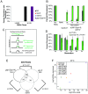
Fig 1. A hypomorphic mutation in gsp-2 results in transgenerational sterility phenotype.
(A) Incidence of males in gsp-2(yp14) was 3.9% at 20°C and increased to 16.8% at 25°C. When L1 animals were shifted to 25°C we saw a similar increase in males (5%, N = 42) to animals grown at 20°C and when L4 animals were shifted incidence of males was 10.7% (N = 49) (B) Progeny of gsp-2(yp14) animals grown at 20°C or 25°C were 6% and 41% Embryonic Lethal, respectively, compared to 97% of gsp-2(tm301) progeny (N = 20). (C-D) gsp-2(yp14) was identified to have a G to A mutation in exon 5 by whole genome sequencing. This results in a D to N amino acid substitution in a well conserved region of GSP-2. (E) When passaged at 20°C for many generations N2, gsp-2(yp14), lab-1 and lab-1; gsp-2(yp14) did not exhibit a loss of transgenerational fertility. (F) gsp-2(yp14) and lab-1 both exhibited loss of fertility at 25°C and were completely sterile by generation F17 and F11 respectively. A double mutant of lab-1;gsp-2 went sterile slightly faster than the individual single mutants and were completely sterile by F10. (N≥40) (G) Analysis of incidence of males showed no jackpots of males at in gsp-2(yp14) animals. *P <.0001 by T-test. Error bars represent standard deviation.
Fig 2
Fig 2. GSP-2 promotes multigenerational transgene silencing.
(A) gsp-2(yp14) mutants do not exhibit single generation RNAi defects while rsd-6 and nrde-2 mutants are defective for single generation RNAi. (B) cpIs12 treated with RNAi remains undetectable for multiple generations after RNAi treatment. However, in gsp-2(yp14);cpIs12 animals treated with RNAi cpIs12 only remains undetectable for one generation and by generation 3 exhibit close to wildtype levels of expression. (C,D) When LP138, a GFP transgene, is passaged as a heterozygote for multiple generations it is silenced in the germline. LP138 passaged as a heterozygote in a gsp-2(yp14) mutants results in only partial silencing over 5 generations suggesting defective heterozygous transgene silencing. (E) Comparison of small RNAs in rsd-6, gsp-2 and spr-5 mutants showing a great overlap in small RNA identity between gsp-2 and spr-5. (F) Graph showing levels of piRNA expression in N2 controls, rsd-6, gsp-2 and spr-5 mutants at both early and late generations grown at 25°C.
Fig 3
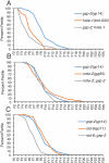
Fig 3. Temperature-sensitive small RNAi mutants exhibited similar times to sterility as gsp-2(yp14) at 25°C.
Germline mortality assays all performed at 25°C (A) Both gsp-2(yp14) and hrde-1 animals exhibit similar times to sterility while gsp-2(yp14);hrde-1 double mutants display a slightly decreased time to sterility. p<.001 (B) gsp-2(yp14), nrde-2 and rsd-6;gsp-2(yp14) animals all go sterile in a similar number of generations. p = .06 (C) gsp-2(yp14) and rsd-6 exhibit similar times to sterility while gsp-2(yp14);rsd-6 double mutants become sterile at a slightly earlier generation. p<.001(N≥40). Significance was tested using a log rank test.
Fig 4
Fig 4. Germline defects occur in gsp-2 and temperature-sensitive small RNA mutants at sterility.
(A-E) Representative images of DAPI stained germlines passaged at 25°C until sterility. The timing of passage differed depending on the genotype as the time to sterility varies (See Fig 3). Germlines of either L4 (A) or adult control and sterile mutant animals were stained, and the germline size quantified as either normal (B), short (C), atrophied (D) or empty (E). (F-G) 6 DAPI bodies in control oocytes (F) and 8 DAPI bodies in gsp-2(yp14) animals (G). (H-I) Germlines from gsp-2(yp14), lab-1, rsd-6, nrde-2, hrde-1, and spr-5 mutants were examined and found to have mostly normal morphology at the L4 stage (H) but exhibited germline atrophy in adult animals (N≥98) (I). (J) In addition to germline atrophy, gsp-2(yp14) animals displayed greater than the wildtype number of 6 DAPI bodies in oocytes at the generation at sterility in 32% of oocytes (N≥100). (K) Quantification of HIM-8 staining showing the % of paired foci for each zone (See S2 Fig) along the germline for all indicated mutants. Error bars represent the standard deviation. P-values were obtained by using a student’s t-test for unpaired samples with unequal variance.
Fig 5
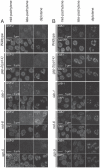
Fig 5. GSP-2 and LAB-1 localization during mid-pachytene to late diplotene.
(A-B) Representative images of nuclei from early pachytene to late diplotene for controls, rsd-6, gsp-2(yp14), spr-5 and lab-1 are stained with an antibody against GSP-2 (A) and an antibody against LAB-1 (B). DAPI images for each nucleus are shown to indicate the specific cell cycle stage. All worms were grown at 25°C for 2 generations and fixed as day 2 adults.
Fig 6
Fig 6. Increased histone phosphorylation is present in gsp-2(yp14) oocytes.
(A-F) Day 2 late stage adults passaged at 25°C stained with an pH3S10 antibody (red) and DAPI marking the DNA (cyan). All samples were prepared at the same time and imaged using identical settings. (A) Wildtype control oocytes show low levels of H3S10p on condensed chromosomes. (B) gsp-2(yp14) oocytes have increased levels of H3S10p covering the entire chromosomes. (C,E, F) lab-1, rsd-6 and hrde-1 mutants also display increased levels of H3S10p but nrde-2 (D) did not. (N≥20) (G-L) Day 2 late generation or sterile adults passaged at 25°C stained with an H3S10p antibody (red) and DAPI marking the DNA (cyan). All samples were prepared at the same time and imaged using identical settings. (G) Control oocytes show low levels of localized H3T3p on the condensed chromosomes. (H) gsp-2(yp14) oocytes contain high levels of H3T3p that are expanded to cover the entire condensed chromosome. (I-L) lab-1, hrde-1, rsd-6 and nrde-2 all display varying levels of increased H3T3p staining compared to wildtype controls. (N≥20) (M) Quantification of fluorescence intensity of H3S10p staining in N2, gsp-2(yp14) animals grown at 20°C and 25°C, rsd-6, nrde-2, hrde-1, and lab-1 shows significant difference in staining intensity between N2 and mutants grown at the same temperature (except for nrde-2) and between gsp-2(yp14) mutants grown at 20°C and 25°C. (N≥20) (N) Quantification of fluorescence intensity of H3T3p staining in N2, gsp-2(yp14) animals grown at 20°C and 25°C, rsd-6, nrde-2, hrde-1, and lab-1 shows significant difference in staining intensity between N2 and mutants grown at the same temperature and between gsp-2(yp14) mutants grown at 20°C and 25°C. (N≥20) Scale bar = 10um.
Fig 7
Fig 7. Temperature-sensitive germline immortality exhibit decreased histone methylation.
(A-B) Control, rsd-6, gsp-2(yp14), spr-5 and lab-1 animals were grown for 2 generations at 25°C and stained for H3K9me2, H3K9me2 and H3K4me3. (A) Images of diakinesis nuclei are shown (B) Intensity measurements for diakinesis nuclei (N≥41). Error bars for all panels indicate standard deviation.
Fig 8
Fig 8. A model for the roles of GSP-2 and small RNA-mediated silencing in promoting germline immortality.
We propose that both GSP-2 and small RNA-mediated silencing regulate the transgenerational inheritance of the epigenome. When these pathways are disrupted loss of epigenetic regulation can lead to germline atrophy. (A) GSP-2 modulates small RNA silencing machinery promoting small RNA silencing potentially through histone dephosphoryation in a manner that promotes epigenetic silencing, (B) Previous work has shown that PRG-1 is important for heterozygous transgene silencing (red = active transgene and black = silenced) in a similar manner to GSP-2. GSP-2/LAB-1 could function to silence small heterozygous regions of DNA, which disrupt meiotic pairing between homologs or cohesion between sister chromatids. This model reflects data presented here and ideas and data from other studies. The model is meant to provoke thoughtful experiments, rather than to represent concepts for which there is definitive experimental proof.

Similar articles

Cited by

References

    1. Sharpless NE, DePinho RA. How stem cells age and why this makes us grow old. Nat Rev Mol Cell Biol. 2007; 703–713. 10.1038/nrm2241 - DOI - PubMed
    1. Ahmed S, Hodgkin J. MRT-2 checkpoint protein is required for germline immortality and telomere replication in C. elegans. Nature. 2000; 159–64. - PubMed
    1. Armanios M, Blackburn EH. The telomere syndromes. Nat Rev Genet. 2012; 693–704. 10.1038/nrg3246 - DOI - PMC - PubMed
    1. Meier B, Clejan I, Liu Y, Lowden M, Gartner A, Hodgkin J, et al. trt-1 is the Caenorhabditis elegans catalytic subunit of telomerase. PLoS Genet. Public Library of Science; 2006; e18. - PMC - PubMed
    1. Bagijn MP, Goldstein LD, Sapetschnig A, Weick EM, Bouasker S, Lehrbach NJ, et al. Function, targets, and evolution of Caenorhabditis elegans piRNAs. Science (80-). 2012; 574–578. - PMC - PubMed

Publication types

MeSH terms

Substances