. 2019 May 28;139(22):2570-2584.
doi: 10.1161/CIRCULATIONAHA.118.036099. Epub 2019 Mar 29.

Endothelial Cells Regulate Physiological Cardiomyocyte Growth via VEGFR2-Mediated Paracrine Signaling

Affiliations

Endothelial Cells Regulate Physiological Cardiomyocyte Growth via VEGFR2-Mediated Paracrine Signaling

Riikka Kivelä et al. Circulation. .

Abstract

Background: Heart failure, which is a major global health problem, is often preceded by pathological cardiac hypertrophy. The expansion of the cardiac vasculature, to maintain adequate supply of oxygen and nutrients, is a key determinant of whether the heart grows in a physiological compensated manner or a pathological decompensated manner. Bidirectional endothelial cell (EC)-cardiomyocyte (CMC) cross talk via cardiokine and angiocrine signaling plays an essential role in the regulation of cardiac growth and homeostasis. Currently, the mechanisms involved in the EC-CMC interaction are not fully understood, and very little is known about the EC-derived signals involved. Understanding how an excess of angiogenesis induces cardiac hypertrophy and how ECs regulate CMC homeostasis could provide novel therapeutic targets for heart failure.

Methods: Genetic mouse models were used to delete vascular endothelial growth factor (VEGF) receptors, adeno-associated viral vectors to transduce the myocardium, and pharmacological inhibitors to block VEGF and ErbB signaling in vivo. Cell culture experiments were used for mechanistic studies, and quantitative polymerase chain reaction, microarrays, ELISA, and immunohistochemistry were used to analyze the cardiac phenotypes.

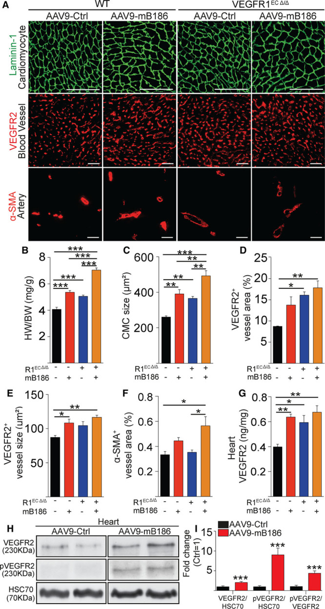
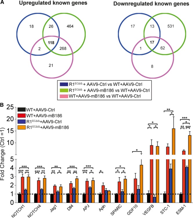
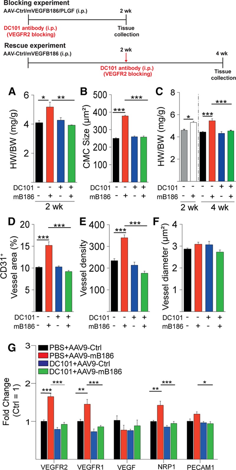
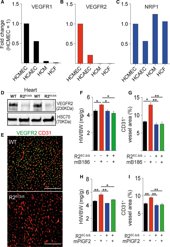
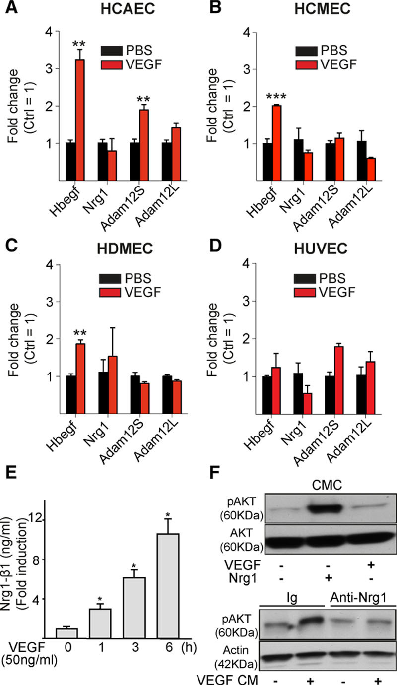
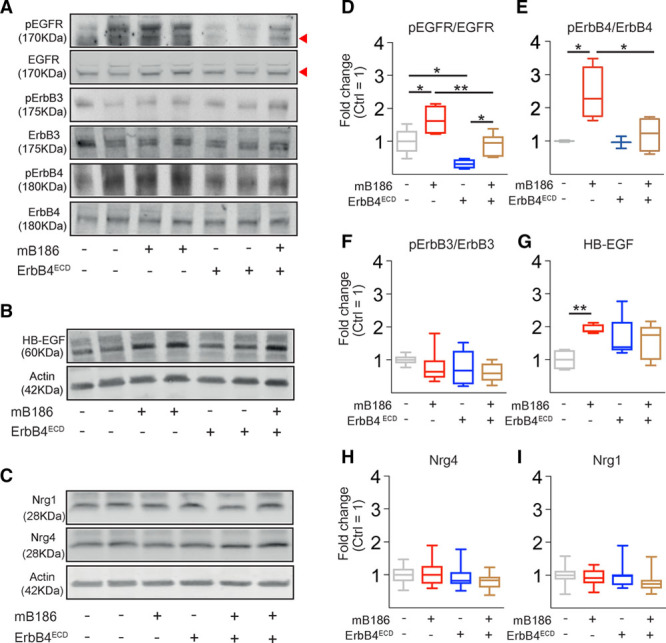
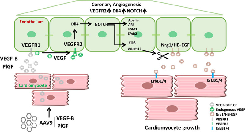
Results: Both EC deletion of VEGF receptor (VEGFR)-1 and adeno-associated viral vector-mediated delivery of the VEGFR1-specific ligands VEGF-B or placental growth factor into the myocardium increased the coronary vasculature and induced CMC hypertrophy in adult mice. The resulting cardiac hypertrophy was physiological, as indicated by preserved cardiac function and exercise capacity and lack of pathological gene activation. These changes were mediated by increased VEGF signaling via endothelial VEGFR2, because the effects of VEGF-B and placental growth factor on both angiogenesis and CMC growth were fully inhibited by treatment with antibodies blocking VEGFR2 or by endothelial deletion of VEGFR2. To identify activated pathways downstream of VEGFR2, whole-genome transcriptomics and secretome analyses were performed, and the Notch and ErbB pathways were shown to be involved in transducing signals for EC-CMC cross talk in response to angiogenesis. Pharmacological or genetic blocking of ErbB signaling also inhibited part of the VEGF-B-induced effects in the heart.

Conclusions: This study reveals that cross talk between the EC VEGFR2 and CMC ErbB signaling pathways coordinates CMC hypertrophy with angiogenesis, contributing to physiological cardiac growth.

Keywords: HB-EGF; VEGF-B; VEGFR-1; angiogenesis; hypertrophy.


Figures

Figure 1.
Figure 1.
VEGFR1 deletion from endothelial cells induces angiogenesis and cardiomyocyte hypertrophy. A, Representative images of staining for cardiomyocytes (Laminin-1), blood vessels (VEGFR2), and arteries (α-SMA) in AAV-VEGFB186–treated or VEGFR1-deleted hearts (R1ECΔ/Δ). B and C, Quantification of heart weight normalized to body weight and cardiomyocyte size (in µm2). D through F, Quantification of blood vessel area, average capillary size (in µm2), and α-SMA area. G, VEGFR2 protein concentration (in ng/mg) in the heart. H, Western blots of total VEGFR2 and phosphorylated VEGFR2 in VEGF-B–overexpressing and wild-type hearts. Heat shock cognate 70 (HSC70) was used as a loading control. I, Quantification of the Western blot signals is shown as fold change compared with AAV-Ctrl treatment. Data are mean±SEM. AAV9 indicates adeno-associated viral vector serotype 9; CMC, cardiomyocyte; Ctrl, control; HW/BW, heart weight/body weight; mB186, mouse VEGF B-186; pVEGFR2, phosphorylated VEGFR2; α-SMA, α-smooth muscle actin; VEGF-B, vascular endothelial growth factor B; VEGFR2, vascular endothelial growth factor receptor 2; and WT, wild type. Two-way ANOVA (Holm-Sidak test) and Student t test were used, as appropriate; *P<0.05, **P<0.01, ***P<0.001 (N=4 per group). Scale bar, 100 µm.
Figure 2.
Figure 2.
Transcriptomic profiling of VEGFR1-deleted or AAV9-VEGFB186–overexpressing adult mouse heart. A, Venn diagram showing the number of common and unique cardiac gene expression changes in hearts deleted of VEGFR1 (R1ECΔ/Δ) or expressing AAV-VEGFB186 or both, compared with control (WT+AAV9-Ctrl) mice. B, Validation of microarray findings for Notch signaling pathway genes and genes identified by secretome analysis in an independent experiment (normalized to Hprt-1). Data are mean±SEM. AAV9 indicates adeno-associated viral vector serotype 9; Ctrl, control; mB186, mouse VEGF B-186; VEGFR2, vascular endothelial growth factor receptor 2; and WT, wild type. Two-way ANOVA with Holm-Sidak multiple comparison test; *P<0.05, **P<0.01, ***P<0.001 (N=4 per group).
Figure 3.
Figure 3.
VEGF-B–induced cardiac hypertrophy and vascular growth are inhibited by blocking VEGFR2 signaling. A and B, Heart weight normalized to body weight (HW/BW; in mg/g) and cardiomyocyte (CMC) size (in µm2) in mice injected with AAV-VEGF-B186 or AAV-Ctrl and treated for 2 weeks with VEGFR2-blocking antibody DC101. In A and B, DC101 was started at the same time as the AAV injections (blocking experiment), and in C, 2 weeks after AAV administration, after hypertrophy had developed (rescue experiment; shown as gray and white bars). These 2 groups were analyzed 2 weeks after AAV and before DC101 treatment was started. D through F, Quantification of blood vessel area, density, and average vessel diameter (blocking experiment). G, Quantification of cardiac mRNAs (normalized to Hprt-1). Data are mean±SEM. AAV indicates adeno-associated viral vector; AAV9, AAV serotype 9; Ctrl, control; mB186 and mVEGFB186, mouse VEGF B-186; NRP1, neuropilin 1; PECAM1, platelet and endothelial cell adhesion molecule 1; PLGF, placental growth factor; VEGF-B, vascular endothelial growth factor B; VEGFR2, vascular endothelial growth factor receptor 2; and WT, wild type. Two-way ANOVA (Holm-Sidak test); *P<0.05, **P<0.01, ***P<0.001 (N= 5 per group).
Figure 4.
Figure 4.
Endothelial VEGFR2 deletion inhibits VEGF-B–induced cardiomyocyte hypertrophy. A through C, Relative VEGFR1, VEGFR2, and NRP1 mRNA expression levels in human coronary microvascular endothelial cells (HCMEC), human coronary arterial endothelial cells (HCAEC), human cardiomyocytes (HCM), and human cardiac fibroblasts (HCF). D and E, VEGFR2 protein levels in R2ECΔ/Δ mouse hearts analyzed by Western blot and immunohistochemistry. Heart to body weight ratio (HW/BW; in mg/g) and blood vessel area (%) are shown in AAV-VEGF-B186 (F and G) and in AAV-PlGF–treated wild-type and R2ECΔ/Δ mice (H and I). Data are mean±SEM. AAV indicates adeno-associated viral vector; HSC70, heat shock cognate 70; mB186, mouse VEGF B-186; mPlGF2, mouse placental growth factor 2; NRP1, neuropilin 1; VEGF-B, vascular endothelial growth factor B; VEGFR1 and VEGFR2, vascular endothelial growth factor receptor 1 and 2; and WT, wild type. Two-way ANOVA (Holm-Sidak test); *P<0.05, **P<0.01, ***P<0.001 (N= 3 per group). Scale bars, 100 µm.
Figure 5.
Figure 5.
VEGF stimulation induces HB-EGF and ADAM12 mRNA expression and Nrg1 release in endothelial cells. A through D, Human cardiac arterial (HCEAC), cardiac microvascular (HCMEC), dermal microvascular (HDMEC), and umbilical vein endothelial cells (HUVEC) were stimulated with vascular endothelial growth factor (VEGF; 100 ng/mL) for 4 hours, and mRNA expression was analyzed. E, Nrg1 levels in conditioned medium from HCMECs stimulated with VEGF. F, Phosphorylation of Akt in cardiomyocytes (CMC) treated with conditioned medium (CM) from HCMECs stimulated with VEGF. Note that Akt activation is blocked with the anti-Nrg1 antibody. HB-EGF indicates heparin-binding epidermal growth factor–like growth factor; Ig, immunoglobulin; and Nrg1, neuregulin 1. Student t test (A through D) and 1-way ANOVA (E); **P<0.01, ***P<0.001 (N=3 per group).
Figure 6.
Figure 6.
Analysis of ErbB ligands and receptors in the AAV-VEGF-B186 and AAV-ErbB4-ECD–treated mouse heart. A through I, Representative Western blots and quantification of phosphorylation of ErbB receptors normalized to total receptors and expression of HB-EGF, Nrg1, and Nrg4 normalized to β-actin (as fold change, Ctrl=1). For epidermal growth factor receptor (EGFR), the lower 175-kDa band was quantified. Numbers per group: pEGFR, n=4–6; pErbB3, n=7–8; pErbB4, n=3–4; HB-EGF, n=5; and Nrg1 and Nrg 4, n=10–15. AAV indicates adeno-associated viral vector; HB-EGF, heparin-binding epidermal growth factor–like growth factor; mB186, mouse VEGF B-186; Nrg1, neuregulin 1; Nrg4, neuregulin 4; pEGFR, phosphorylated epidermal growth factor receptor; and pErbB3 and pErbB4, phosphorylated ErbB3 and ErbB4. Shapiro-Wilk normality test and Mann–Whitney multiple comparison test; *P<0.05, **P<0.01.
Figure 7.
Figure 7.
VEGF-B–induced changes in signaling molecules related to cardiac hypertrophy are inhibited by both VEGFR2 and ErbB blocking. A and B, VEGF-B–induced increased transcript expression of PI3K-p110β, Akt, Carp/Ankrd1, and Tbx3 and decreased expression of C/EBPβ in the heart were blocked by both DC101 and afatinib treatment. C through F, Afatinib restored the VEGF-B–induced phosphorylation of Akt and Erk, with a similar trend for S6 kinase (S6K). Data are mean±SEM. AAV9 indicates adeno-associated viral vector serotype 9; Ctrl, control; mB186, mouse VEGF B-186; pErk1/2, phosphorylated Erk1/2; PI3K, phosphoinositide 3-kinase; pS6K, phosphorylated S6 kinase; and VEGF-B, vascular endothelial growth factor B. Two-way ANOVA (Holm-Sidak test); *P<0.05, **P<0.01 (N=4–5 per group).
Figure 8.
Figure 8.
Schematic illustrating endothelial cell to cardiomyocyte cross talk in angiogenesis-induced cardiomyocyte hypertrophy. Adeno-associated viral vector serotype 9 (AAV9) encoding vascular endothelial growth factor B (VEGF-B) or placental growth factor (PlGF) transduces the cardiomyocytes. The secreted VEGF-B and PlGF bind to vascular endothelial growth factor receptor 1 (VEGFR1) in the endothelial cells and increase the bioavailability of endogenous vascular endothelial growth factor (VEGF) to VEGF receptor 2 (VEGFR2). Activation of VEGFR2 in endothelial cells induces the activation of Dll4/NOTCH signaling, which leads to coronary angiogenesis and arteriogenesis. In addition, endothelial cell VEGFR2 activation upregulates the expression of Id1, apelin, APJ, ESM1, EfnB2, Klk8, and Adam12. Adam12 and Klk8–mediated shedding of heparin-binding epidermal growth factor–like growth factor (HB-EGF) and neuregulin 1 (Nrg1) from the endothelial cell surface produces soluble cleaved forms of these proteins. HB-EGF and Nrg1 bind and activate epidermal growth factor receptor 1 (EGFR1 [ErbB1]) and ErbB4 in cardiomyocytes and promote cardiomyocyte growth.

Comment in

References

    1. van Berlo JH, Maillet M, Molkentin JD. Signaling effectors underlying pathologic growth and remodeling of the heart. J Clin Invest. 2013;123:37–45. doi: 10.1172/JCI62839. - PMC - PubMed
    1. Mann N, Rosenzweig A. Can exercise teach us how to treat heart disease? Circulation. 2012;126:2625–2635. doi: 10.1161/CIRCULATIONAHA.111.060376. - PMC - PubMed
    1. Walsh K, Shiojima I. Cardiac growth and angiogenesis coordinated by intertissue interactions. J Clin Invest. 2007;117:3176–3179. doi: 10.1172/JCI34126. - PMC - PubMed
    1. Hudlicka O, Brown M, Egginton S. Angiogenesis in skeletal and cardiac muscle. Physiol Rev. 1992;72:369–417. doi: 10.1152/physrev.1992.72.2.369. - PubMed
    1. Shimizu I, Minamino T. Physiological and pathological cardiac hypertrophy. J Mol Cell Cardiol. 2016;97:245–262. doi: 10.1016/j.yjmcc.2016.06.001. - PubMed

Publication types

MeSH terms

Substances