Case Reports
. 2019 Aug;25(4):447-453.
doi: 10.1177/1591019919830767. Epub 2019 Mar 28.

Cerebral foreign body reaction due to hydrophilic polymer embolization following aneurysm treatment by pipeline flow diversion device

Affiliations
Case Reports

Cerebral foreign body reaction due to hydrophilic polymer embolization following aneurysm treatment by pipeline flow diversion device

Thomas Rand Geisbush et al. Interv Neuroradiol. 2019 Aug.

Abstract

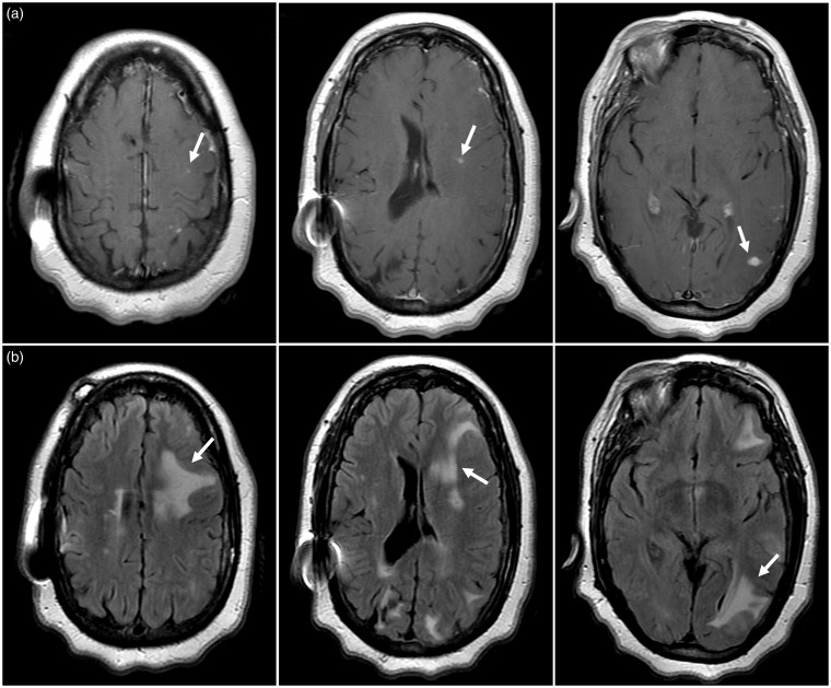
The use of flow diverting stents for wide based, intracranial aneurysms has become an invaluable treatment option. While intracranial hemorrhage and ischemic stroke from dislodged atherosclerotic emboli are common adverse events, the potential for delayed granulomatous inflammation from possible hydrophilic polymer emboli is rarely recognized. We present a unique case in which visible chipping of the pusher wire for stent placement was observed, followed by clinical and radiographic evidence suggestive of a delayed foreign body reaction to intracranial hydrophilic polymer emboli. A 55-year-old woman underwent placement of a Pipeline embolization device for a left-sided, broad-based aneurysm at the base of the internal carotid artery and posterior communicating artery. Two months later she developed right-sided focal neurological deficits. Imaging showed ipsilateral focal edema and enhancing lesions with contrast. Although not confirmed with biopsy and histopathology, clinical and radiographic evidence suggests that this patient probably experienced a delayed foreign body reaction to hydrophilic polymer emboli from compromised procedural equipment during flow diverting stent placement. Although previously described, this is the first instance to our knowledge in whichvisible chipping of the pusher wire was observed on a Pipeline embolization device.

Keywords: Granulomatous inflammation; cerebral aneurysm; flow diverting stent; foreign body reaction; hydrophilic polymer embolization.


Figures

Figure 1.
Figure 1.
Digital subtraction angiography (DSA) following left internal carotid artery (ICA) injection during Pipeline embolization device (PED) placement. DSA in the anteroposterior (a) and lateral (b) projections demonstrate an 8 mm × 6 mm aneurysm with a 7.6 mm neck arising near the origin of the posterior communicating artery (a, b thick arrows). (c) A non-digital subtraction angiogram of the PED stent shortly after deployment crossing the span of the aneurysm base (single bracket). Distal to the stent is the guidewire tip coil (solid thin arrow) and proximal to the stent is the proximal marker/pusher wire (dashed thin arrow).
Figure 2.
Figure 2.
Brain magnetic resonance imaging findings following new neurological symptoms 2 months after Pipeline embolization device (PED) treatment. (a) Axial post-contrast T1-weighted images show multiple small enhancing foci (arrows) within the left cerebral hemisphere. (b) Axial fluid-attenuated inversion recovery (FLAIR) images identify increased signal abnormality surrounding the regions of contrast enhancement that is most consistent with vasogenic edema (arrows). Regions of FLAIR signal abnormality in the right cerebral hemisphere represent baseline non-specific white matter disease based on comparison with pre-PED treatment neuroimaging studies. Regions of T2 signal abnormality in the right cerebral hemisphere did not demonstrate associated enhancement and were felt to be most consistent with non-specific periventricular white matter changes.
Figure 3.
Figure 3.
Digital subtraction angiography (DSA) following new symptom development 2 months after Pipeline embolization device (PED) placement. DSA in the anteroposterior (a) and lateral (b) projections demonstrate minimal residual filling (arrows) of the left posterior communicating artery aneurysm previously treated by flow diversion. No other vascular abnormality is identified to account for the patient's symptoms.
Figure 4.
Figure 4.
Brain magnetic resonance imaging (MRI) after treatment for presumed hydrophilic polymer emboli (HPE) using steroids. MRI was obtained 2 weeks after treatment with oral steroids, and she was symptom free at the time of this MRI. (a) Axial post-contrast T1-weighted images show an interval reduction in the number and conspicuity of small enhancing foci (arrows) within the left cerebral hemisphere. (b) Axial fluid-attenuated inversion recovery (FLAIR) images demonstrate an interval reduction in the degree of edema surrounding the enhancing foci following steroid treatment (arrows).

References

    1. Mehta RI, Mehta RI. Polymer-induced central nervous system complications following vascular procedures: spectrum of iatrogenic injuries and review of outcomes. Hum Pathol 2016; 53: 178–190. - PMC - PubMed
    1. Shapiro M, Ollenschleger MD, Baccin C, et al. Foreign body emboli following cerebrovascular interventions: clinical, radiographic, and histopathologic features. Am J Neuroradiol 2015; 36: 2121–2126. - PMC - PubMed
    1. Lorentzen AO, Nome T, Bakke SJ, et al. Cerebral foreign body reaction after carotid aneurysm stenting. Interv Neuroradiol 2016; 22: 53–57. - PMC - PubMed
    1. Briganti F, Leone G, Marseglia M, et al. Endovascular treatment of cerebral aneurysms using flow-diverter devices: a systematic review. Neuroradiol J 2015; 28: 365–375. - PMC - PubMed
    1. Becske T, Kallmes DF, Saataci I, et al. Pipeline for uncoilable or failed aneurysms: results from a multicenter clinical trial. Radiology 2013; 267: 858–868. - PubMed

Publication types