

Risk factors for upper adjacent segment degeneration after multi-level posterior lumbar spinal fusion surgery
- PMID: 30922408
- PMCID: PMC6437868
- DOI: 10.1186/s13018-019-1126-9
Risk factors for upper adjacent segment degeneration after multi-level posterior lumbar spinal fusion surgery
Abstract
Background: Posterior lumbar spinal fusion has been widely used in degenerative lumbar stenosis, but adjacent segment degeneration (ASD) was common. Researchers have found many risk factors for ASD after one or two levels of surgery, but few clinical studies focused on multi-level surgery. The purpose of this study was to clarify risk factors for upper ASD after multi-level posterior lumbar spinal fusion.
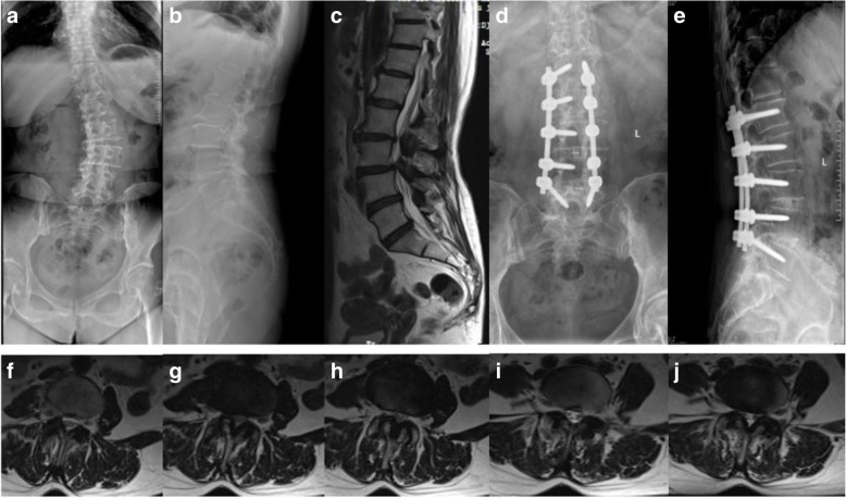
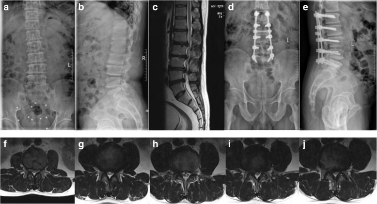
Methods: A retrospective study was performed on the clinical data of 71 patients with degenerative lumbar stenosis who underwent multi-level (at least 3 levels) posterior lumbar spinal fusion from January 2013 to December 2016. Two groups were divided according to lamina and posterior ligamentous complex (PLC) maintenance of proximal fixed vertebrae in surgery. In the 22 patients of group A, the proximal fixed vertebral lamina and PLC were not resected, and in the 49 patients of group B, the proximal fixed vertebral lamina and PLC were resected completely. Age, sex, body mass index (BMI), number of fixed vertebrae and fused levels, spinopelvic parameters, coronal Cobb angle, and modified Pfirrmann grading system were measured for each patient. A Cox proportional hazards model was used to analyze risk factors for upper ASD.
Results: No symptomatic ASD was found during the follow-up period. Patients who underwent proximal fixed vertebral lamina and PLC resection had a significantly higher percentage of radiographic ASD (P = 0.042). The Cox proportional hazards model showed that age, sex, BMI, preoperative lumbar lordosis, sacral slope, pelvic tilt, coronal Cobb angle, number of fixed vertebrae, and interbody fusion levels had no significant differences for radiographic ASD. But a preoperative modified Pfirrmann grade higher than 3, a high degree of preoperative pelvic incidence, and more decompressed levels had statistical significance (P = 0.024, 0.041, and 0.008, respectively).
Conclusions: A preoperative modified Pfirrmann grade higher than 3, a high degree of preoperative pelvic incidence, and more decompressed levels might be risk factors for upper radiographic ASD after multi-level posterior lumbar spinal fusion surgery.
Keywords: Adjacent segment degeneration; Decompression; Disc degeneration; Multi-level; Pelvic incidence; Posterior lumbar fusion; Risk factor.
Conflict of interest statement
Authors’ information
Not applicable.
Ethics approval and consent to participate
This retrospective study was approved by the Institutional Ethics Board of The First Affiliated Hospital of Zhengzhou University. All enrolled patients were informed and agreed to provide relevant data for this study. The methods were carried out in accordance with the relevant guidelines.
Consent for publication
Not applicable.
Competing interests
The authors declare that they have no competing interests.
Publisher’s Note
Springer Nature remains neutral with regard to jurisdictional claims in published maps and institutional affiliations.
Figures
References
MeSH terms
LinkOut - more resources
Full Text Sources
Medical
