. 2019 Jul;27(7):1064-1074.
doi: 10.1016/j.joca.2019.03.003. Epub 2019 Mar 25.

Mechanical loading inhibits cartilage inflammatory signalling via an HDAC6 and IFT-dependent mechanism regulating primary cilia elongation

Affiliations

Mechanical loading inhibits cartilage inflammatory signalling via an HDAC6 and IFT-dependent mechanism regulating primary cilia elongation

S Fu et al. Osteoarthritis Cartilage. 2019 Jul.

Abstract

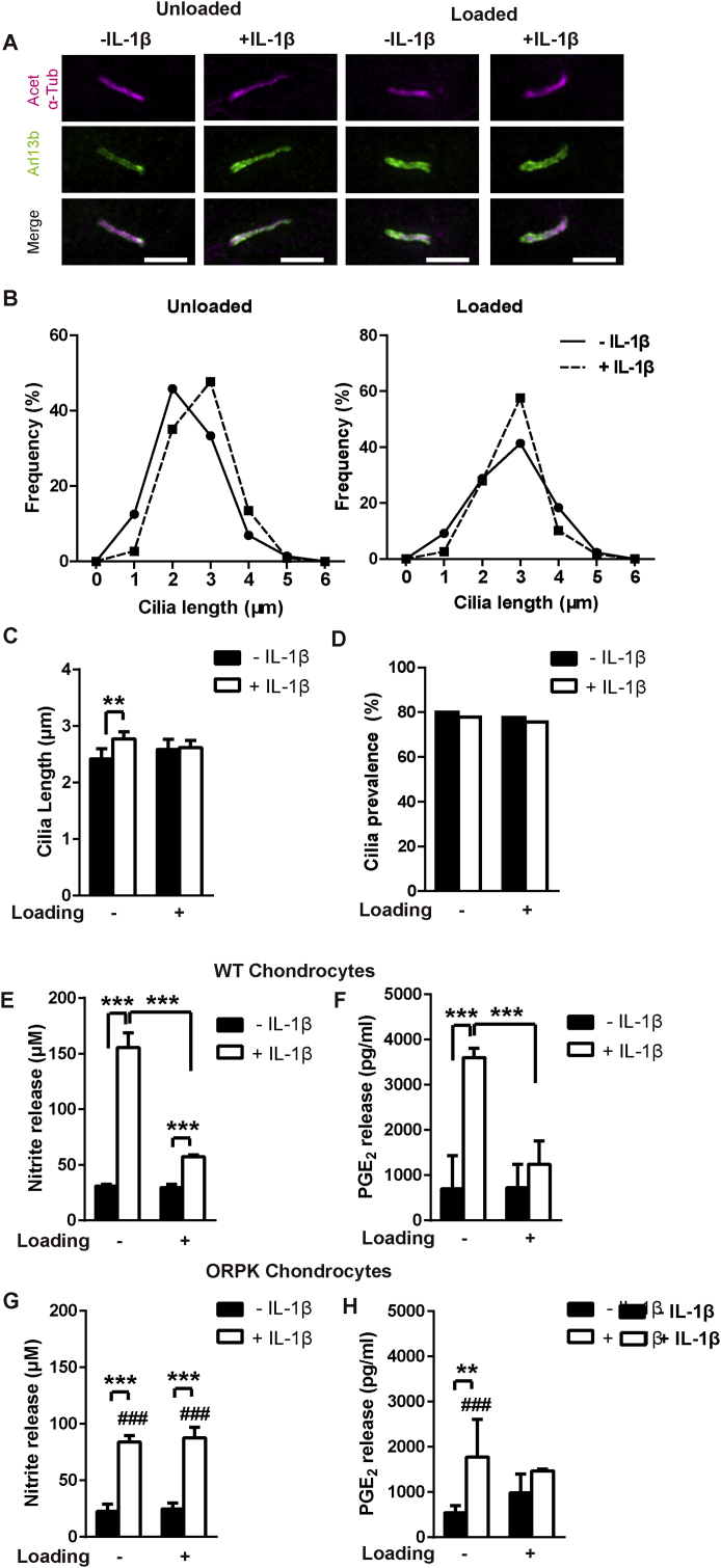
Objective: Physiological mechanical loading reduces inflammatory signalling in numerous cell types including articular chondrocytes however the mechanism responsible remains unclear. This study investigates the role of chondrocyte primary cilia and associated intraflagellar transport (IFT) in the mechanical regulation of interleukin-1β (IL-1β) signalling.

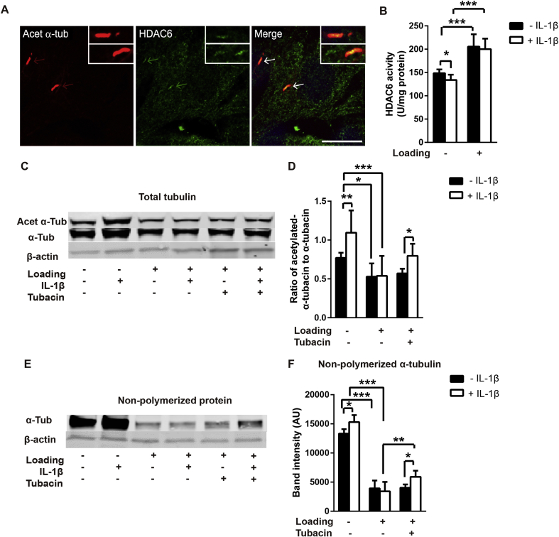
Design: Isolated chondrocytes and cartilage explants were subjected to cyclic mechanical loading in the presence and absence of the cytokine IL-1β. Nitric oxide (NO) and prostaglandin E2 (PGE2) release were used to monitor IL-1β signalling whilst Sulphated glycosaminoglycan (sGAG) release provided measurement of cartilage degradation. Measurements were made of HDAC6 activity and tubulin polymerisation and acetylation. Effects on primary cilia were monitored by confocal and super resolution microscopy. Involvement of IFT was analysed using ORPK cells with hypomorphic mutation of IFT88.

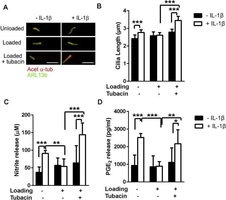
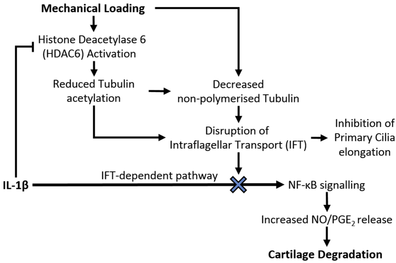
Results: Mechanical loading suppressed NO and PGE2 release and prevented cartilage degradation. Loading activated HDAC6 and disrupted tubulin acetylation and cilia elongation induced by IL-1β. HDAC6 inhibition with tubacin blocked the anti-inflammatory effects of loading and restored tubulin acetylation and cilia elongation. Hypomorphic mutation of IFT88 reduced IL-1β signalling and abolished the anti-inflammatory effects of loading indicating the mechanism is IFT-dependent. Loading reduced the pool of non-polymerised tubulin which was replicated by taxol which also mimicked the anti-inflammatory effects of mechanical loading and prevented cilia elongation.

Conclusions: This study reveals that mechanical loading suppresses inflammatory signalling, partially dependent on IFT, by activation of HDAC6 and post transcriptional modulation of tubulin.

Keywords: Chondrocyte; HDAC6; IFT; IL-1β; Primary cilia; Tubulin.


Figures

Fig. 1
Fig. 1
Mechanical loading inhibits IL-1β signalling and cartilage degradation. (A) NO (nitrite) release and (B) PGE2 release were measured in the culture media (samples n = 6, from donors N = 2) for primary chondrocytes ± IL-1β (10 ng/ml) in the presence or absence of cyclic tensile strain (CTS, 0–10%, 0.33 Hz, 24 h). (C) NO release and (D) Sulphated glycosaminoglycan (sGAG) release were measured in the media (n = 6, N = 2) for full depth cartilage explants ± IL-1β (10 ng/ml) in the presence or absence of cyclic compressive strain (CCS, 0–10%, 0.33 Hz, 24 h). Statistical differences based on Two-way ANOVA with Bonferroni's post hoc test.
Fig. 2
Fig. 2
Mechanical loading blocks IL-1β induced elongation of chondrocyte primary cilia indicating disruption to intraflagellar transport (IFT) which is required for inflammatory signalling. (A) Representative SIM maximum intensity projection images of chondrocyte primary cilia ± IL-1β (1 ng/ml) in the presence or absence of mechanical loading in the form of cyclic tensile strain (CTS, 0–10%, 0.33 Hz, 24 h). Cilia were labelled for acetylated-α-tubulin (Acet-α-Tub, far-red) and arl13b (green). Scale bar represents 2 μm. (B) Frequency histograms of cilia length in unloaded and loaded cells and (C) corresponding mean cilia length (n ≈ 80–100 cilia) and (D) cilia prevalence (n ≈ 100 cells). Statistical analyses based on Mann–Whitney test for cilia length and Chi-square test for cilia prevalence. Changes in (E, G) nitrite and (F, H) PGE2 release for wild type (WT) (E, F) and ORPK (G, H) chondrocytes ± IL-1β (1 ng/ml) in the presence or absence of cyclic tensile strain (CTS, 0–10%, 0.33 Hz, 24 h, n = 4, N = 2). Statistical analyses based on two-way ANOVA with Bonferroni's post hoc test; # indicates significant difference between WT and ORPK.
Fig. 3
Fig. 3
Mechanical loading blocks IL-1β induced tubulin acetylation and reduces non-polymerized tubulin expression via activation of HDAC6. (A) Representative confocal immunofluorescence for acetylated-α-tubulin (Acet-α-Tub, red), HDAC6 (green) in cultured chondrocytes showing co-localization on the cilia axoneme. (B) HDAC6 activity for cells ± IL-1β (1 ng/ml) in the presence or absence of cyclic tensile strain (CTS, 0–10%, 0.33 Hz, 24 h, n = 6, N = 2). Western blot analysis of tubulin based on (C–D) total protein levels and (E–F) non-polymerized protein for cells ± IL-1β (1 ng/ml) in the presence or absence of cyclic tensile strain (CTS) as above. (C and E) Representative blots and corresponding semi-quantitative analysis of (D) ratio of acetylated-α-tubulin to α-tubulin and (F) non-polymerized α-tubulin expression (n = 6). Statistical analyses based on two-way ANOVA with Bonferroni's post hoc test.
Fig. 4
Fig. 4
Reduction in non-polymerized tubulin by taxol treatment disrupts IFT-mediated cilia elongation and inhibits IL-1β signalling. Chondrocytes were cultured for 24 hrs with and without IL-1β (1 ng/ml) and taxol (15 μM). Corresponding measurement of (A) primary cilia length (n ≈ 100 cilia); and (B) prevalence (n ≈ 100 cells) and the release of the pro-inflammatory mediators, (C) NO and (D) PGE2 (n = 6). Statistical analyses based on Mann–Whitney test for cilia length, Chi-square test for cilia prevalence and two-way ANOVA with Bonferroni's post hoc test for NO and PGE2 release.
Fig. 5
Fig. 5
HDAC6 mediates the mechanical suppression of IL-1β-induced cilia elongation and inflammatory signalling. Chondrocytes were cultured on Flexcell membranes ± IL-1β (1 ng/ml), ± cyclic tensile strain (CTS, 0–10%, 0.33 Hz, 24 h) in the presence or absence of tubacin (0.5 μM). (A) Representative confocal immunofluorescence maximum intensity projection images of primary cilia labelled for acetylated-α-tubulin (Acet-α-Tub, red) and arl13b (green). Corresponding data for (B) cilia length (n ≈ 60–80 cilia), (C) NO release and (D) PGE2 release (n = 6, N = 2). Statistical analyses based on Kruskal Wallis test for cilia length and two-way ANOVA with Bonferroni's post hoc test for NO and PGE2 release.
Fig. 6
Fig. 6
In unloaded cells, tubacin prevents IL-1β-mediated tubulin acetylation, cilia elongation and nitrite release. (A) Cilia length (n ≈ 80–100 cilia) and (B) nitrite release (n = 6) for chondrocytes cultured in 0.2 μM, 0.5 μM, 1 μM or 2 μM tubacin ± IL-1β (1 ng/ml). Statistical analyses based on Mann–Whitney test for cilia length, one-way ANOVA with Bonferroni's post hoc test for NO release; * demonstrates statistical significance compared with untreated cells; # compared with cells treated with IL-1β in the absence of tubacin. (C–E) Western blot analysis of tubulin based on total protein levels for cells ± IL-1β (1 ng/ml) and ±tubacin (0.5 μM). (C) Representative blot and semi-quantitative analysis of (D) acetylated-α-tubulin and (E) α-tubulin expression and (F) the corresponding acetylation ratio (n = 3). Statistical analyses based on two-way ANOVA with Bonferroni's post hoc test.
Fig. 7
Fig. 7
Proposed mechanism through which physiological mechanical loading suppresses IL-1β-induced NO/PGE2 production and downstream cartilage degradation. We suggest that the anti-inflammatory effects of mechanical loading are mediated by mechanosensitive HDAC6 activation resulting in disruption of tubulin acetylation and a reduction in the availability of non-polymerized tubulin, which disrupt IFT and associated primary cilia elongation. This mechanical modulation of IFT therefore suppresses IFT-dependent IL-1β signalling events in the form of NO and PGE2 release, thereby reducing cartilage degradation.

References

    1. Goetz S.C., Anderson K.V. The primary cilium: a signalling centre during vertebrate development. Nat Rev Genet. 2010;11:331. - PMC - PubMed
    1. Muhammad H., Rais Y., Miosge N., Ornan E.M. The primary cilium as a dual sensor of mechanochemical signals in chondrocytes. Cell Mol Life Sci. 2012;69:2101–2107. - PMC - PubMed
    1. Singla V., Reiter J.F. The primary cilium as the cell's antenna: signaling at a sensory organelle. Science. 2006;313:629–633. - PubMed
    1. Wann A., Knight M. Primary cilia elongation in response to interleukin-1 mediates the inflammatory response. Cell Mol Life Sci. 2012;69:2967–2977. - PMC - PubMed
    1. Dinsmore C., Reiter J.F. Endothelial primary cilia inhibit atherosclerosis. EMBO Rep. 2016;17:156–166. - PMC - PubMed

Publication types