. 2019 Apr;40(4):745-753.
doi: 10.3174/ajnr.A6016. Epub 2019 Mar 28.

Spontaneous Intracranial Hypotension: A Systematic Imaging Approach for CSF Leak Localization and Management Based on MRI and Digital Subtraction Myelography

Affiliations

Spontaneous Intracranial Hypotension: A Systematic Imaging Approach for CSF Leak Localization and Management Based on MRI and Digital Subtraction Myelography

R I Farb et al. AJNR Am J Neuroradiol. 2019 Apr.

Abstract

Background and purpose: Localization of the culprit CSF leak in patients with spontaneous intracranial hypotension can be difficult and is inconsistently achieved. We present a high yield systematic imaging strategy using brain and spine MRI combined with digital subtraction myelography for CSF leak localization.

Materials and methods: During a 2-year period, patients with spontaneous intracranial hypotension at our institution underwent MR imaging to determine the presence or absence of a spinal longitudinal extradural collection. Digital subtraction myelography was then performed in patients positive for spinal longitudinal extradural CSF collection primarily in the prone position and in patients negative for spinal longitudinal extradural CSF collection in the lateral decubitus positions.

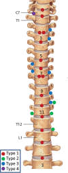
Results: Thirty-one consecutive patients with spontaneous intracranial hypotension were included. The site of CSF leakage was definitively located in 27 (87%). Of these, 21 were positive for spinal longitudinal extradural CSF collection and categorized as having a ventral (type 1, fifteen [48%]) or lateral dural tear (type 2; four [13%]). Ten patients were negative for spinal longitudinal extradural CSF collection and were categorized as having a CSF-venous fistula (type 3, seven [23%]) or distal nerve root sleeve leak (type 4, one [3%]). The locations of leakage of 2 patients positive for spinal longitudinal extradural CSF collection remain undefined due to resolution of spontaneous intracranial hypotension before repeat digital subtraction myelography. In 2 (7%) patients negative for spinal longitudinal extradural CSF collection, the site of leakage could not be localized. Nine of 21 (43%) patients positive for spinal longitudinal extradural CSF collection were treated successfully with an epidural blood patch, and 12 required an operation. Of the 10 patients negative for spinal longitudinal extradural CSF collection (8 localized), none were effectively treated with an epidural blood patch, and all have undergone (n = 7) or are awaiting (n = 1) an operation.

Conclusions: Patients positive for spinal longitudinal extradural CSF collection are best positioned prone for digital subtraction myelography and may warrant additional attempts at a directed epidural blood patch. Patients negative for spinal longitudinal extradural CSF collection are best evaluated in the decubitus positions to reveal a CSF-venous fistula, common in this population. Patients with CSF-venous fistula may forgo further epidural blood patch treatment and go on to surgical repair.


Figures

Fig 1.
Fig 1.
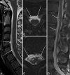
Spinal longitudinal extradural collections. A, Sagittal T2 FSE. B, Reformatted axial T2 SPACE images show SLECs (arrows) and displaced dura outlined by the CSF. C and D, Images similar to A and B of the same patient show similar findings in the lower thoracic region.
Fig 2.
Fig 2.
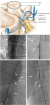
Type 1 CSF leak (SLEC-P). A, Schematic drawing shows the relationship of the intervertebral disc spur and a ventral dural tear. B, “Shoot though” lateral subtracted image of the thoracic spine DSM with the patient positioned prone on the table. The patient's head is toward the top of the image and feet at the bottom. The contrast material can be seen escaping from the ventral aspect of the thecal sac at the T7–8 level (arrow).
Fig 3.
Fig 3.
Type 2 CSF leak (SLEC-P). A, Schematic depiction of a proximal nerve root sleeve tear bridging the epidural and neural foraminal compartments. B–G, From a single patient. B, Sagittal T1WI of the brain shows the engorged pituitary gland (open white arrow) and dural thickening on the clivus (short white arrows). C, Sagittal T1WI of the brain shows a “positive venous distension sign” with a convex undersurface of the middle third of the dominant transverse sinus (short black arrow). D, T2-weighted axial MR image of the thoracic spine shows SLECs (white arrows) external to the dura (white arrowhead). E, Subtracted image from a prone thoracic DSM shows a posterolateral collection of contrast (black arrow). F and G, Subtracted and nonsubtracted images from a repeat right lateral decubitus DSM show contrast leaking into the extradural space (black arrows) from a tear along the proximal aspect of the right T11 root sleeve (long white arrow). Note the BB (nipple marker) placed on the skin for landmarking (dashed white arrow).
Fig 4.
Fig 4.
Type 3 CSF leak (SLEC-N). A, Schematic depiction of a CSF-to-venous fistula arising from a dural tear along the nerve root sleeve beyond the epidural compartment (see text). Nonsubtracted (B) and magnified, subtracted (C) images from separate left-side-down DSM runs in a patient negative for SLEC with SIH. A small vascular structure, in keeping with a tortuous vein of a CVF, can be seen coursing away from the root sleeve (arrows). An incidental normal diverticulum is also noted at the level above (arrowhead). D and E, Nonsubtracted images of decubitus DSMs of 2 other similarly presenting patients negative for SLEC demonstrating CVFs. Globular collections of contrast (dashed arrow) are commonly seen near the expected zone of origin of the vein, possibly representing a focal extravasation (pseudomeningocele) of contrast or a diverticulum from which the vein appears to arise.
Fig 5.
Fig 5.
Type 4 CSF leak (SLEC-N). A, Schematic depiction of a distal nerve root sleeve dural tear occurring beyond the epidural compartment extravasating into the surrounding fascial planes and loose connective tissue without loculation or fistulization. B, CT of the head. Sagittal reformat in a patient negative for SLEC demonstrates large low-density (bilateral) subdural hemorrhages (asterisk). Note the prominent “venous distension sign” (short arrow) despite the large subdural hemorrhages. C, Axial CT image obtained 10–20 minutes post-DSM shows subtle extravasated contrast in the region of the right C8 nerve (arrow). Note that on this nondynamic CT (slightly degraded due to beam-hardening artifacts associated with the shoulders), there is little to help distinguish this extravasated contrast from a normal diverticulum. D, Subtracted image from a right-side-down decubitus DSM shows extravasation of contrast (arrows) into the paraspinal tissues from a leak along the mid-to-distal right C8 nerve root sleeve.
Fig 6.
Fig 6.
Distribution of CSF leaks.

References

    1. Kranz PG, Gray L, Amrhein TJ. Spontaneous intracranial hypotension: 10 myths and misperceptions. Headache 2018;58:948–59 10.1111/head.13328 - DOI - PubMed
    1. Kranz PG, Malinzak MD, Amrhein TJ, et al. . Update on the diagnosis and treatment of spontaneous intracranial hypotension. Curr Pain Headache Rep 2017;21:37 10.1007/s11916-017-0639-3 - DOI - PubMed
    1. Schievink WI. Spontaneous spinal cerebrospinal fluid leaks. Cephalalgia 2008;28:1345–56 10.1111/j.1468-2982.2008.01776.x - DOI - PubMed
    1. Schievink WI, Schwartz MS, Maya MM, et al. . Lack of causal association between spontaneous intracranial hypotension and cranial cerebrospinal fluid leaks. J Neurosurg 2012;116:749–54 10.3171/2011.12.JNS111474 - DOI - PubMed
    1. Schievink WI, Maya MM, Jean-Pierre S, et al. . A classification system of spontaneous spinal CSF leaks. Neurology 2016;87:673–79 10.1212/WNL.0000000000002986 - DOI - PubMed

LinkOut - more resources