Review
. 2019 May;20(5):298-313.
doi: 10.1038/s41583-019-0152-2.

Emerging connections between cerebellar development, behaviour and complex brain disorders

Affiliations
Review

Emerging connections between cerebellar development, behaviour and complex brain disorders

Aaron Sathyanesan et al. Nat Rev Neurosci. 2019 May.

Abstract

The human cerebellum has a protracted developmental timeline compared with the neocortex, expanding the window of vulnerability to neurological disorders. As the cerebellum is critical for motor behaviour, it is not surprising that most neurodevelopmental disorders share motor deficits as a common sequela. However, evidence gathered since the late 1980s suggests that the cerebellum is involved in motor and non-motor function, including cognition and emotion. More recently, evidence indicates that major neurodevelopmental disorders such as intellectual disability, autism spectrum disorder, attention-deficit hyperactivity disorder and Down syndrome have potential links to abnormal cerebellar development. Out of recent findings from clinical and preclinical studies, the concept of the 'cerebellar connectome' has emerged that can be used as a framework to link the role of cerebellar development to human behaviour, disease states and the design of better therapeutic strategies.


Conflict of interest statement

Competing interests policy There is NO Competing Interest.

Figures

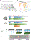
Figure 1.
Figure 1.. Essential features of cerebellar connections, circuitry and development
(a) General scheme of input and output connections to and from the cerebellum. Main inputs include spinal cord, inferior olive and pontine nucleus. Main outputs include connections from cerebellar nuclei to cerebral cortex via the thalamus. (b) Cellular anatomy and circuit connections within the cerebellar cortex. PCs are shown in orange, GCs in magenta, MLIs in purple, GoCs in green, unipolar brush cell interneuron in cyan, input from inferior olive is shown in blue, input from the brain and spinal cord is shown in grey (c) When the human and rodent timelines are aligned based on major cellular/developmental events in the cerebellum, in humans the window of vulnerability to injury (indicated by red numbers) is mostly late gestational, while in preclinical rodent models, it is mostly postnatal. (d) Cellular schematic of events depicted in the timeline in panel ‘a’ showing EGL expansion (grey), dendritic arborization (PCs blue), and white matter interneuron migration in the 1st postnatal week. Migration of GCs into the IGL (green) continues in the second postnatal week with concomitant reduction of EGL, and circuit formation. In the adult, cerebellar circuitry formation is completed, the EGL has disappeared, and MLIs (salmon) have been integrated into the cerebellar cortical circuitry.
Figure 2.
Figure 2.. Features of the cerebellar connectome and dependent factors
(a) Cerebellar connectome developmental trajectories can be plotted along three dimensions – space (gold), time (turquoise), and information (purple). (b) Spatial factors include cell density and regional volumes (defined by y1 and y2), and effective distance of secreted molecules (defined by y3). Temporal factors include time of onset of developmental events such as interneuron migration (Δt), and information factors include cell-to-cell communication events such as spontaneous PC firing, and long range communication between PCs and cortical neurons. Cerebellar development is a protracted process, making this brain region particularly vulnerable to a broad range of abnormalities. This early vulnerability is represented by the probability space defined as a function of spatial, temporal and information factors (red box). While trajectories may start out as typically-developing (solid blue trajectory, TD), alterations in the developmental disruption probability space can cause altered paths resulting in overconnected connectomes (short space dashed trajectory, e.g. high functioning autism), underconnected connectomes (long space dashed trajectory, e.g. low functioning autism), or hypoplasticity (dotted trajectory, e.g. Down syndrome).
Figure 3.
Figure 3.. Regions of cerebellar developmental vulnerability and multisensory integration.
(a) Certain cerebellar regions are more vulnerable to abnormalities than others in the domain of complex disorders. Structural MRI images are from figure 2 in Ref. . Images on the left show voxels (red) with significant structural changes associated with autism spectrum disorders, compared to typically developing controls. Images on the right are corresponding sections with functional connectivity map as described in Ref. . Colors denote networks, namely, blue – somatomotor, green – dorsal attention, violet – ventral attention, cream – limbic, orange – frontoparietal, red – default network (b) Granule cells (GCs) (grey) integrate sensory information (auditory, visual and somatosensory) resulting in multisensory integration. Golgi cells (GoCs, green) are also involved in modulation of sensory information flowing into the cerebellum. Integration of sensory information from complex stimuli such as speech is impaired in autism spectrum disorder.
Figure 4.
Figure 4.. Deep brain stimulation of the cerebellum in mouse.
(a) Schematic illustrating the experimental set-up for stimulating cerebellar circuits in adult mice (b) Schematic of the tissue section (bottom) illustrates the targeting of the stimulating electrodes to the interposed nuclei (In), with reference to the overlying cortex (CCtx) and adjacent cerebellar nuclei. The fourth ventricle is labeled as 4V (c) Schematics illustrating a deep brain stimulation approach of the cerebellar circuit in a mouse model of dystonia. Mobility is immediately improved after stimulation begins (DBS on condition). Adapted with permission from Ref .

References

    1. Stoodley CJ, Valera EM & Schmahmann JD Functional topography of the cerebellum for motor and cognitive tasks: an fMRI study. NeuroImage 59, 1560–1570, doi:10.1016/j.neuroimage.2011.08.065 (2012). - DOI - PMC - PubMed
    1. Buckner RL The cerebellum and cognitive function: 25 years of insight from anatomy and neuroimaging. Neuron 80, 807–815, doi:10.1016/j.neuron.2013.10.044 (2013). - DOI - PubMed
    1. Timmann D et al. The human cerebellum contributes to motor, emotional and cognitive associative learning. A review. Cortex; a journal devoted to the study of the nervous system and behavior 46, 845–857, doi:10.1016/j.cortex.2009.06.009 (2010). - DOI - PubMed
    1. Strick PL, Dum RP & Fiez JA Cerebellum and nonmotor function. Annual review of neuroscience 32, 413–434, doi:10.1146/annurev.neuro.31.060407.125606 (2009). - DOI - PubMed
    1. Piaget J The origin of intelligence in the child. (1953).

Publication types