Review
. 2019 Mar 16:13:16.
doi: 10.1186/s13037-019-0196-2. eCollection 2019.

Outcomes after surgical treatment of acetabular fractures: a review

Affiliations
Review

Outcomes after surgical treatment of acetabular fractures: a review

Navid Ziran et al. Patient Saf Surg. .

Abstract

Acetabular fractures are fractures that extend into the hip joint and pose a challenge for orthopaedic trauma surgeons. The first known descriptions of surgical fixation of acetabular fractures were case reports in 1943. In 1964, Robert Judet, Jean Judet, and Émile Letournel published a landmark article describing a classification system and surgical approaches to treat acetabular fractures. These teachings had a significant effect on clinical outcomes after surgical fixation of acetabular fractures. In 1980, Letournel demonstrated 80% good-to-excellent results in 492 hips, and in 2012, Joel Matta demonstrated 79% survivorship in 816 patients follow surgical acetabular fixation. Both Letournel and Matta have definitively shown that anatomic reduction of the fracture is the most influential factor predictive of clinical outcome. The intent of this review is to summarize the salient factors affecting clinical outcomes after surgical treatment of acetabular fractures.

Keywords: Acetabular fracture; Hip joint; Post-traumatic arthritis; Surgical fixation; Survivorship.


Conflict of interest statement

Not applicable.Not applicable.The authors declare that they have no competing interests.Springer Nature remains neutral with regard to jurisdictional claims in published maps and institutional affiliations.

Figures

Fig. 1
Fig. 1
Anteroposterior (a), obturator oblique (b), and iliac oblique (c) view of a T-shaped fracture showing displacement of the femoral head and inadequate roof arc measurement. Surgery is indicated. Permission was granted for utilization of this figure in this review
Fig. 2
Fig. 2
Anteroposterior (a), obturator oblique (b), and iliac oblique (c) view of a T-shaped fracture showing congruence of the femoral head out of traction and adequate roof-arc measurements. Closed treatment is indicated. Permission was granted for utilization of this figure in this review
Fig. 3
Fig. 3
a Bone-density mapping of 5 mm of subchdral bone in an intact right acetabulum couresy of Lubovsky et al. 2013. The highest bone density is in the superior and posterior portions of the acetabulum. Injury to these regions tends to correlate with worse prognosis. Density map of a fractured left acetabulum is shown in panel b. Fracture lines are marked as yellow circles and a bony defect is shown in dark blue. Permission was granted for figure utilization in this review
Fig. 4
Fig. 4
Line drawing and (A, b) and anteroposterior (B), iliac oblique (C) radiographs of the “gull-sign.” The fragment is a partial fracture of the posterior column (A) not including the ischial tuberosity; it involves the posterior portion of the quadrilateral surface, the ischial spine, and the roof is hinged inwards. This displacement of the posterior quadrilateral surface manifests as a re-duplication of the ilioischial line and the rotated roof segment, adjacent to the intact roof, appears as a gull in flight. Permission was granted for utilization of this figure in this review
Fig. 5
Fig. 5
Pre-operative (ac), immediate post-operative (d), and 21-year post-operative radiographs (eh) of a patient after reduction and fixation of a both-column acetabular fracture. At 21 years, the left hip superior joint space is intact and patient has a good clinical outcome
Fig. 6
Fig. 6
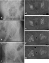
Pre-operative (ac) radiographs and CT (dg) of a patient who sustained a comminuted transtectal transverse acetabular fracture with involvement of the posterior wall
Fig. 7
Fig. 7
Post-operative AP pelvis (a) and Judet view (bc) radiographs of a transtectal + posterior wall acetabular fracture after limited open reduction internal fixation and acute total hip arthroplasty

References

    1. Judet R, Judet J, Letournel É. Fractures of the acetabulum: classification and surgical approaches for open reduction. Preliminary report. J Bone Joint Surg Am. 1964;46:1615–1646. doi: 10.2106/00004623-196446080-00001. - DOI - PubMed
    1. Letournel É. Acetabulum fractures: classification and management. Clin Orthop Relat Res. 1980;151:81–106. - PubMed
    1. Tannast MM, Najibi SS, Matta JM. Two to twenty-year survivorship of the hip in 810 patients with operatively treated acetabular fractures. J Bone Joint Surg Am. 2012;94(17):1559–1567. doi: 10.2106/JBJS.K.00444. - DOI - PubMed
    1. Letournel E, Judet R. Fracture of the acetabulum. 2nd edition. Berlin: Springer-Verlag; 1993.
    1. Levine MA. Treatment of central fractures of the acetabulum. A case report. J Bone and Joint Surg. 1943;25:902–906.