. 2019 Jul 11;21(7):878-889.
doi: 10.1093/neuonc/noz058.

EZHIP/CXorf67 mimics K27M mutated oncohistones and functions as an intrinsic inhibitor of PRC2 function in aggressive posterior fossa ependymoma

Affiliations

EZHIP/CXorf67 mimics K27M mutated oncohistones and functions as an intrinsic inhibitor of PRC2 function in aggressive posterior fossa ependymoma

Jens-Martin Hübner et al. Neuro Oncol. .

Abstract

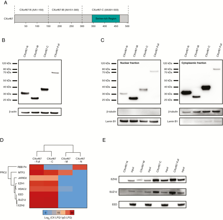
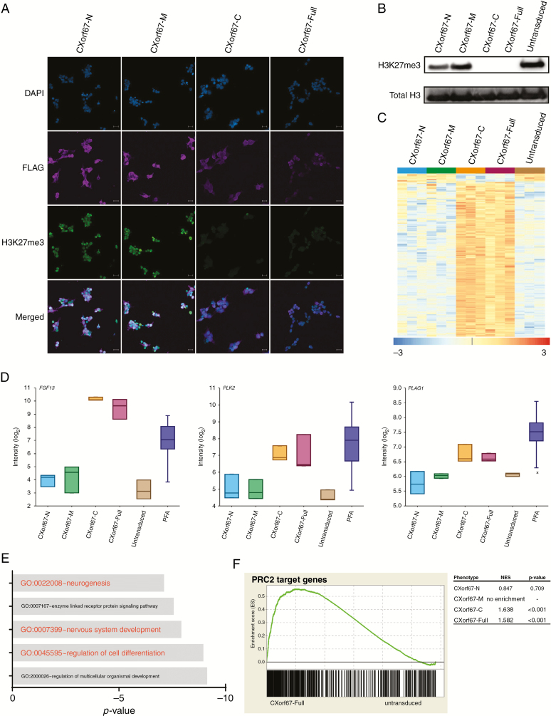
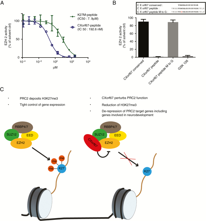
Background: Posterior fossa A (PFA) ependymomas are one of 9 molecular groups of ependymoma. PFA tumors are mainly diagnosed in infants and young children, show a poor prognosis, and are characterized by a lack of the repressive histone H3 lysine 27 trimethylation (H3K27me3) mark. Recently, we reported overexpression of chromosome X open reading frame 67 (CXorf67) as a hallmark of PFA ependymoma and showed that CXorf67 can interact with enhancer of zeste homolog 2 (EZH2), thereby inhibiting polycomb repressive complex 2 (PRC2), but the mechanism of action remained unclear.

Methods: We performed mass spectrometry and peptide modeling analyses to identify the functional domain of CXorf67 responsible for binding and inhibition of EZH2. Our findings were validated by immunocytochemistry, western blot, and methyltransferase assays.

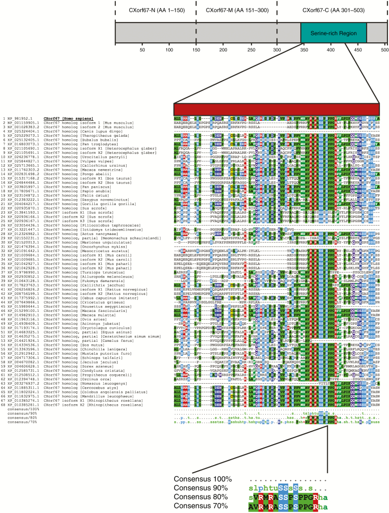
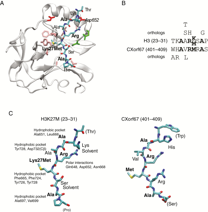
Results: We find that the inhibitory mechanism of CXorf67 is similar to diffuse midline gliomas harboring H3K27M mutations. A small, highly conserved peptide sequence located in the C-terminal region of CXorf67 mimics the sequence of K27M mutated histones and binds to the SET domain (Su(var)3-9/enhancer-of-zeste/trithorax) of EZH2. This interaction blocks EZH2 methyltransferase activity and inhibits PRC2 function, causing de-repression of PRC2 target genes, including genes involved in neurodevelopment.

Conclusions: Expression of CXorf67 is an oncogenic mechanism that drives H3K27 hypomethylation in PFA tumors by mimicking K27M mutated histones. Disrupting the interaction between CXorf67 and EZH2 may serve as a novel targeted therapy for PFA tumors but also for other tumors that overexpress CXorf67. Based on its function, we have renamed CXorf67 as "EZH Inhibitory Protein" (EZHIP).


Figures

Fig. 1
Fig. 1
Mass spectrometry analysis of CXorf67 truncates reveals interaction of the C-terminus of CXorf67 with PRC2 core components. (A) Overview of CXorf67 and the 3 truncates used in this study. The serine-rich region of the protein is highlighted in turquoise. (B) Detection of FLAG-tagged CXorf67 truncates and the full-length protein in lysates of HEK293T cells after transduction. (C) Western blot analysis of nuclear and cytoplasmic fractions obtained from transduced HEK293T cells. Both fractions contain CXorf67 truncates and the full-length protein. Lamin B1 was used as a nuclear loading control and β-tubulin as a loading control for the cytoplasmic fraction. (D) Heatmap depicting PRC2 components interacting with CXorf67-Full and at least 1 truncate. The scale bar depicts the ratio of the log2 signal intensities (LFQ) of PRC2 related proteins within each CXorf67 variant experiment over their respective immunoglobulin G control. (E) Validation of CXorf67-C interaction with EZH2 and SUZ12 by co-IP. A faint interaction can also be observed for CXorf67-M.
Fig. 2
Fig. 2
The C-terminus of CXorf67 is crucial for PRC2 binding and inhibition. (A) Staining of HEK293T cells expressing CXorf67 truncates or the full-length protein using an H3K27me3 antibody. Cells expressing CXorf67-C or CXorf67-Full show a reduced H3K27me3 signal. Scale bar: 20 µm. (B) Western blot analysis of H3K27me3 levels in HEK293T cells expressing CXorf67 truncates or the full-length protein. A strong downregulation of H3K27me3 can be observed for CXorf67-C and CXorf67-Full. (C) Heatmap depicting significantly (P < 0.05) deregulated genes (n = 198) upon overexpression of CXorf67-Full in HEK293T cells. A strong deregulation of the same genes can also be observed for CXorf67-C expressing cells (n = 3 per condition). The scale bar depicts the z-scores of the gene expression levels. (D) Comparison of expression levels for FGF13, PLK2, and PLAG1 in transduced cell lines (n = 3), untransduced control cells (n = 3), and PFA tumors (n = 55). (E) GO enrichment analysis of CXorf67-Full upregulated genes (n = 188). X-axis depicts the p-value (log10). (F) GSEA showing significant enrichment of PRC2 target genes in CXorf67-C and CXorf67-Full cells compared with untransduced control cells. No significant enrichment is observed for the other truncated variants. A representative plot for CXorf67-Full is shown. NES: normalized enrichment score.
Fig. 3
Fig. 3
The C-terminus of human CXorf67 contains a highly conserved amino acid sequence. Multiple sequence alignment of CXorf67 ortholog protein sequences identifies a highly conserved 13 amino acid sequence in the C-terminus of human CXorf67. Alignments were performed for the entire protein but the figure shows only the indicated region in the C-terminus of CXorf67. Amino acids that match the human CXorf67 sequence are colored.
Fig. 4
Fig. 4
CXorf67 inhibits PRC2 via a small, highly conserved peptide sequence located in its C-terminus. (A) Structure of EZH2 bound to H3K27M where the bound peptide is shown as sticks and colored according to atom type. Residues in EZH2 binding to the peptide are shown as sticks and colored according to residue type: hydrophobic, pink; polar, green; negative, red. (B) Alignment of the peptide regions from H3 and CXorf67, where conserved residues are in bold. Residues above and below the alignment are those identified as allowed substitutions by perusing all orthologs of the 2 proteins in eggNOG. (C) The 2 peptides in the same orientation, where part of the CXorf67 peptide was modeled into the EZH2 structure using Modeller. The relevant binding pocket residues in EZH2 are labeled for each residue in H3K27M and conserved residues are shown in boldface. Structures were created using visual molecular dynamics.
Fig. 5
Fig. 5
A CXorf67 peptide incorporating the conserved region potently inhibits EZH2. (A) Methyltransferase assay to evaluate the inhibitory capabilities of the CXorf67 and the K27M peptides. (B) Evaluation of EZH2 inhibition by CXorf67 peptide variants at a single concentration of 10 µM. Peptide variants encompass the conserved region alone (CXorf67 conserved), the longer variant incorporating surrounding amino acids (CXorf67 peptide) and the longer peptide variant in which the methionine has been replaced by a glycine (CXorf67 peptide M to G). GSK126 was used as a positive control. (C) Proposed model of CXorf67-mediated H3K27 hypomethylation. When CXorf67 is absent, PRC2 mediates trimethylation of H3K27, resulting in a tight regulation of gene expression. In contrast, the presence of CXorf67 results in the inhibition of PRC2. In detail, the conserved domain of CXorf67 sequesters EZH2, thereby abolishing its methyltransferase activity. The resulting decrease in H3K27me3 levels allows the de-repression of PRC2 target genes, including genes involved in neurodevelopment which contributes to the tumorigenesis of PFA ependymoma.

References

    1. Ostrom QT, de Blank PM, Kruchko C, et al. . Alex’s lemonade stand foundation infant and childhood primary brain and central nervous system tumors diagnosed in the United States in 2007–2011. Neuro Oncol. 2015;16(Suppl 10):x1–x36. - PMC - PubMed
    1. Kilday JP, Rahman R, Dyer S, et al. . Pediatric ependymoma: biological perspectives. Mol Cancer Res. 2009;7(6):765–786. - PubMed
    1. Pajtler KW, Witt H, Sill M, et al. . Molecular classification of ependymal tumors across all CNS compartments, histopathological grades, and age groups. Cancer Cell. 2015;27(5):728–743. - PMC - PubMed
    1. Witt H, Mack SC, Ryzhova M, et al. . Delineation of two clinically and molecularly distinct subgroups of posterior fossa ependymoma. Cancer Cell. 2011;20(2):143–157. - PMC - PubMed
    1. Wani K, Armstrong TS, Vera-Bolanos E, et al. ; Collaborative Ependymoma Research Network A prognostic gene expression signature in infratentorial ependymoma. Acta Neuropathol. 2012;123(5):727–738. - PMC - PubMed

Publication types

MeSH terms