. 2019 Mar 28;6(1):14.
doi: 10.1186/s40634-019-0180-x.

Effect on inclined medial proximal tibial articulation for varus alignment in advanced knee osteoarthritis

Affiliations

Effect on inclined medial proximal tibial articulation for varus alignment in advanced knee osteoarthritis

Tomoharu Mochizuki et al. J Exp Orthop. .

Abstract

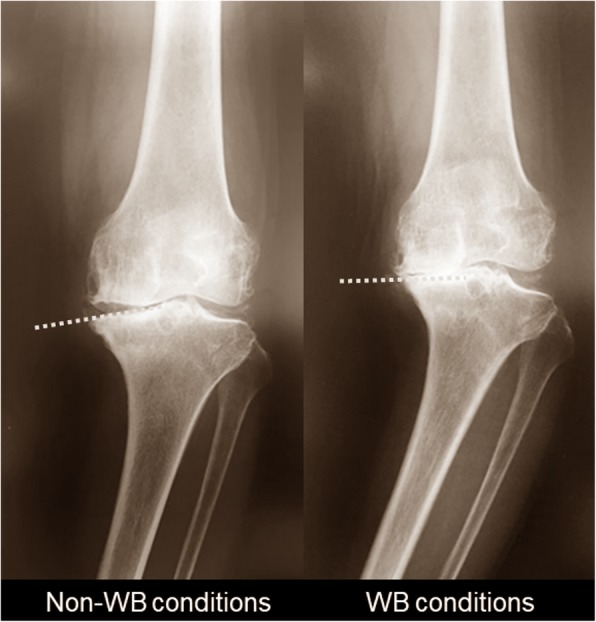
Background: The inclination of the medial compartment of the proximal tibia (MCT) is assumed to be a critical factor for varus alignment in advanced knee osteoarthritis (OA). This study was aimed at investigating; (1) whether the inclination of MCT is aligned parallel to the ground under weight-bearing (WB) conditions; (2) whether this is associated with the change in alignment and the relative position between the bones; and (3) whether the tibia or femur mainly contributes to the changes.

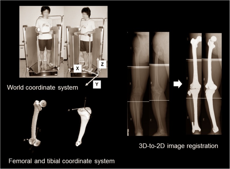
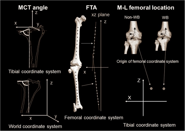
Methods: We examined 102 knees (84 women, 18 men; mean 75 years). A three-dimensional (3D) assessment system was applied on biplanar whole lower extremity radiographies using 3D-to-2D image registration technique. The evaluation parameters were 1) MCT angle, 2) femorotibial angle (FTA), 3) medial-lateral femoral location to the tibia (M-L femoral location), 4) WB line passing point, and 5) tibial position to WB line (tibial position) and 6) femoral postion to WB line (femoral position). Each parameter was evaluated in non-WB and WB conditions, and the differences (Δ-parameters).

Results: MCT angle in the world coordinate system was larger than that in the tibial coordinate system (p < 0.0001). ΔMCT angle was correlated with ΔFTA (p = 0.002) and ΔM-L femoral location (p = 0.004). The tibial position was the more dominant factor for ΔMCT angle (p = 0.001), ΔFTA (p < 0.0001), and ΔWB line passing point (p < 0.0001) .

Conclusions: The inclination in MCT was aligned parallel to the ground under WB conditions (tibial parallel phenomenon). The parallel phenomenon was associated with the change of alignment and the relative position between the bones in the coronal plane. These phenomena were produced mainly by the tibia, not the femur.

Level of evidence: Level IV.

Keywords: Inclination in the medial compartment of the proximal tibia; Knee osteoarthritis; Tibial parallel phenomenon; Varus alignment; Weight-bearing conditions.


Conflict of interest statement

Ethics approval and consent to participate

The study was approved by the Institutional Review Board in Niigata University. Appropriate consents, permissions and releases were obtained.

Consent for publication

All presentations had consent to publish.

Competing interests

The authors did not receive and will not receive any benefits or funding from any commercial party related directly or indirectly to the subject of this article.

Publisher’s Note

Springer Nature remains neutral with regard to jurisdictional claims in published maps and institutional affiliations.

Figures

Fig. 1
Fig. 1
Knee radiographs in advanced knee OA under non-WB and WB conditions. An articular surface in the MCT under WB conditions was aligned parallel to the ground, compared to that under non-WB conditions
Fig. 2
Fig. 2
Schematic diagram showing the varus alignment mechanism in advanced knee OA. As the hypothesis, in a coronal plane, a tibial parallel phenomenon may produce varus malalignment and the position change between the femur and tibia under WB conditions in advanced knee OA
Fig. 3
Fig. 3
The 3D lower extremity alignment assessment system under WB conditions based on biplanar long lower extremity radiographs, applying the 3D-to-2D image registration techniques. 3D bone models and the anatomic coordinate system of the femur and tibia were reconstructed. The 3D bone model included the definitions of the parameters in the bony morphology and whole lower extremity alignment. To assess the 3D parameters “in the standing position” under WB conditions, biplanar long lower extremity radiographs were obtained in the standing position. Applying the 3D-to-2D image registration technique, projected outline points of each 3D model were the finite edge points of the 2D shadow created from the projections of all visible triangular surfaces of the 3D model. To precisely assess the position and angle to the ground, the world coordinate system was reconstructed
Fig. 4
Fig. 4
Schematic diagram showing the MCT angle, FTA, and M–L femoral location. MCT angle under non-WB conditions in the tibial coordinate system was defined as the angle between the tangential line of an articular surface in MCT and the z-axis of the tibial coordinate system. As the assessment plane, the coronal plane passing the tibial coordinate system origin point was used. MCT angle under WB conditions in the world coordinate system was defined as the angle between the tangential line of an articular surface in the MCT and the z-axis of the world coordinate system. As the assessment plane, the coronal plane passing the middle section between the anterior- and posterior-most points of the medial compartment in the world coordinate system was used. In terms of FTA, the anatomic longitudinal axes were defined as a regression line obtained from approximating distances from these 10 centroids in the femur and 12 centroids in the tibia by the least squares method, respectively. FTA was assessed as the angle between the anatomic longitudinal axes of the femur and tibia in the coronal plane of the femoral coordinate system. Regarding M–L femoral location, the M–L location of the femur relative to the tibia was defined as the location of the origin point of the femoral coordinate system, and was assessed in the coronal plane of the tibial coordinate system
Fig. 5
Fig. 5
Schematic diagram showing the WB line passing point, tibial position, and femoral position. Regarding WB line passing point, WB line was defined as the line connecting the femoral head with the center of the ankle joint in a 3D space. WB line passing point was the location of the WB line on the xy plane in the tibial coordinate system. The WB line in the tibial coordinate system was described by percent indication (0% = origin in the tibial coordinate system, + 100% = medial-most point in MCT, and ≥ 100% = passing point outside from MCT). The tibial position was defined as the angle between the WB line and the x-axis in the tibial coordinate system. The femoral position was defined as the angle between the WB line and the surgical epicondylar axis in the tibial coordinate system

References

    1. Aberg AC, Thorstensson A, Tarassova O, Halvorsen K. Calculations of mechanisms for balance control during narrow and single-leg standing in fit older adults: a reliability study. Gait Posture. 2011;34(3):352–357. doi: 10.1016/j.gaitpost.2011.05.025. - DOI - PubMed
    1. Akamatsu Y, Mitsugi N, Taki N, Takeuchi R, Saito T. Relationship between low bone mineral density and varus deformity in postmenopausal women with knee osteoarthritis. J Rheumatol. 2009;36(3):592–597. doi: 10.3899/jrheum.080699. - DOI - PubMed
    1. Ariumi A, Sato T, Kobayashi K, Koga Y, Omori G, Minato I, Endo N. Three-dimensional lower extremity alignment in the weight-bearing standing position in healthy elderly subjects. J Orthop Sci. 2010;15(1):64–70. doi: 10.1007/s00776-009-1414-z. - DOI - PubMed
    1. Birmingham TB, Hunt MA, Jones IC, Jenkyn TR, Giffin JR. Test–retest reliability of the peak knee adduction moment during walking in patients with medial compartment knee osteoarthritis. Arthritis Rheum. 2007;57(6):1012–1017. doi: 10.1002/art.22899. - DOI - PubMed
    1. Canny A. A computational approach to edge detection. IEEE Transactions on Pattern Analysis and Machine Intelligence PAMI. 1986;8(6):679–698. doi: 10.1109/TPAMI.1986.4767851. - DOI - PubMed